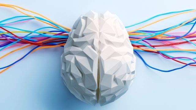
A study in zebrafish shows that neuronal microexons regulate brain activity by modulating cAMP signaling. Loss of these fragments leads to hyperactivity and sleep disruption, offering insights into mechanisms linked to neurodevelopmental disorders.
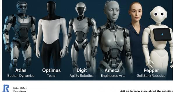
Humanoid robots are rapidly evolving, demonstrating significant advancements in capabilities and applications across various industries. A detailed comparison among the most prominent models available today reveals significant distinctions and areas of specialization, each catering to different sectors and operational requirements. Here is a comparison table for humanoid robots in the market.
Researchers from the University of Colorado Boulder, CU Anschutz, and Colorado State University have developed a set of experimental treatments that may help aging and damaged joints repair themselves in a matter of weeks. The therapies have shown promising results in animal studies, where they reversed signs of osteoarthritis and restored joint health.
The new approaches include a regenerative injection designed to be administered directly into a joint, as well as a biomaterial-based repair system that encourages the body’s own cells to rebuild damaged cartilage.
The work recently received a major boost from the federal Advanced Research Projects Agency for Health (ARPA-H), which announced that the team will move forward to the next stage of a project worth up to $33.5 million. The research is part of the ARPA-H Novel Innovations for Tissue Regeneration in Osteoarthritis (NITRO) program, led by ARPA-H Program Manager Dr. Ross Uhrich.

Can biology answer questions that once belonged only to philosophy?
Alex Rosenberg argues that Darwinian biology transformed not only science but also our understanding of morality, meaning, mind, and human purpose, bringing traditionally philosophical questions into the scientific domain.
0:00 What Is the Philosophy of Biology 1:14 How Darwin Changed the Nature of Inquiry 4:27 How Philosophers Help Biologists 6:48 Biology and the Philosophy of Mind 9:43 Can Biology Answer Philosophy’s Biggest Questions.
Alexander Rosenberg is an American philosopher and novelist. He is the R. Taylor Cole Professor of Philosophy at Duke University, well known for contributions to philosophy of biology and philosophy of economics. Rosenberg describes himself as a \.
A study led by researchers at Children’s Hospital of Philadelphia (CHOP) demonstrates a new method of using decellularized cartilage with patient-specific cells to help enlarge pediatric airways narrowed as a result of severe subglottic stenosis. Researchers demonstrate that this new method is faster, more effective and able to overcome issues associated with the current standard grafts, such as donor site morbidity, insufficient tissue volume and a delayed timeline. The findings are published in the journal Nature Communications.
Severe subglottic stenosis is a narrowing of the airway below the vocal cords and above the trachea. An estimated 20,000 infants per year are affected by this condition. The most severe cases require laryngotracheal reconstruction (LTR), an open-airway surgery used to enlarge the airway by implanting cartilage taken from the rib cage.
While LTR is used to successfully treat thousands of children with subglottic stenosis, in many cases, young children lack enough costal cartilage—the cartilage connecting the ribs to the sternum—for these grafts. As a result, operations often need to be delayed, leaving the child attached to a tracheostomy tube until they are older, and there is a higher risk of needing follow-up surgery because the airway is at risk of narrowing again.
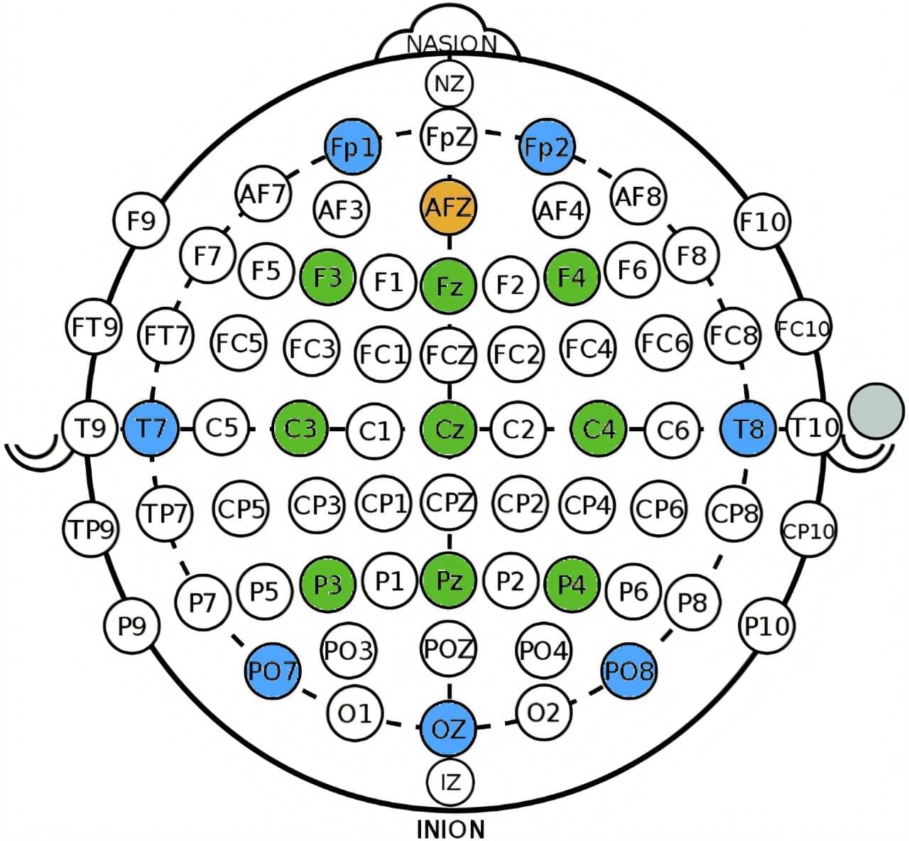
A new approach for identifying signs of hidden awareness in people who cannot speak or move after severe brain injury has been demonstrated by researchers at the University of Bath in the U.K.
The system detects patterns of brain activity through a wearable headset using an advanced application of brain-computer interface (BCI) technology.
Across multiple experimental sessions, the researchers uncovered signs of consciousness that were previously undetected in unresponsive patients.
The first human trial of epigenetic reprogramming is underway.
Scientists are testing whether three genes can make old cells behave like young ones again while avoiding the cancer risk that has challenged the field for years.