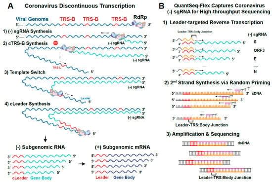
Coronaviruses use discontinuous transcription to generate subgenomic RNAs (sgRNAs) that encode structural and accessory proteins. However, the factors regulating sgRNA abundance in SARS-CoV-2 remain unclear. Here, we combined strand-specific RNA sequencing, RNA–RNA interaction mapping, prediction of RNA folding energies, and targeted mutagenesis to define the regulation of (–) sgRNA synthesis in SARS-CoV-2 infection. We demonstrated that the relative (–) sgRNA abundance across viral genes is stable throughout infection and largely correlates with corresponding (+) sgmRNA levels. Through meta-analysis of published SPLASH data, we found that the frequency of long-range interactions between the 5′ genomic transcription regulatory sequence TRS-Leader and downstream TRS-Body sequences correlates with sgRNA abundance.
Category: mapping
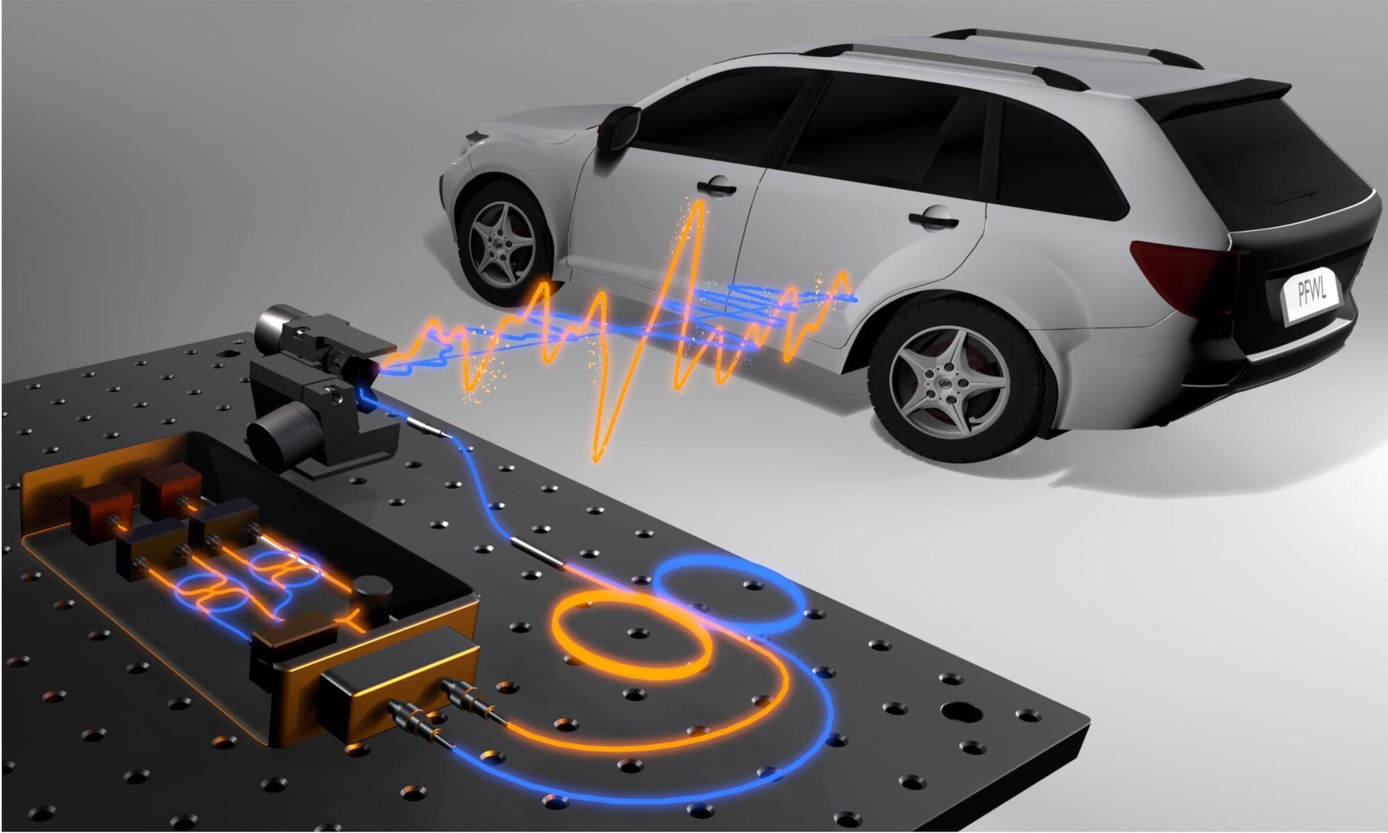
New lidar system maps location, speed and material properties in a single measurement
Researchers have developed a new kind of lidar system that simultaneously measures the location, speed and material properties of objects in a scene. This type of information could be useful for applications such as robotics, autonomous driving and remote sensing.
Lidar uses laser pulses to measure distances and create highly detailed 3D maps of objects and terrain. However, most commercial lidar systems, such as those used in autonomous cars, primarily measure distance.
“Although some emerging lidar technologies can also measure velocity, real-world perception often requires understanding an object’s surface as well,” said Dongyu Du from the University of Toronto in Canada. “Our new system uses a single measurement at each scanned point to capture millimeter-accurate distance, velocity and surface material while using eye-safe laser power.”

The Neuroscience of Happiness and Pleasure
The evolutionary imperatives of survival and procreation, and their associated rewards, are driving life as most animals know it. Perhaps uniquely, humans are able to consciously experience these pleasures and even contemplate the elusive prospect of happiness. The advanced human ability to consciously predict and anticipate the outcome of choices and actions confers on our species an evolutionary advantage, but this is a double-edged sword, as John Steinbeck pointed out as he wrote of “the tragic miracle of consciousness” and how our “species is not set, has not jelled, but is still in a state of becoming” (Steinbeck and Ricketts 1941). While consciousness allows us to experience pleasures, desires, and perhaps even happiness, this is always accompanied by the certainty of the end.
Nevertheless, while life may ultimately meet a tragic end, one could argue that if this is as good as it gets, we might as well enjoy the ride and in particular to maximize happiness. Yet, it is also true that for many happiness is a rare companion due to the competing influences of anxiety and depression.
In order to help understand happiness and alleviate the suffering, neuroscientists and psychologists have started to investigate the brain states associated with happiness components and to consider the relation to well-being. While happiness is in principle difficult to define and study, psychologists have made substantial progress in mapping its empirical features, and neuroscientists have made comparable progress in investigating the functional neuroanatomy of pleasure, which contributes importantly to happiness and is central to our sense of well-being.
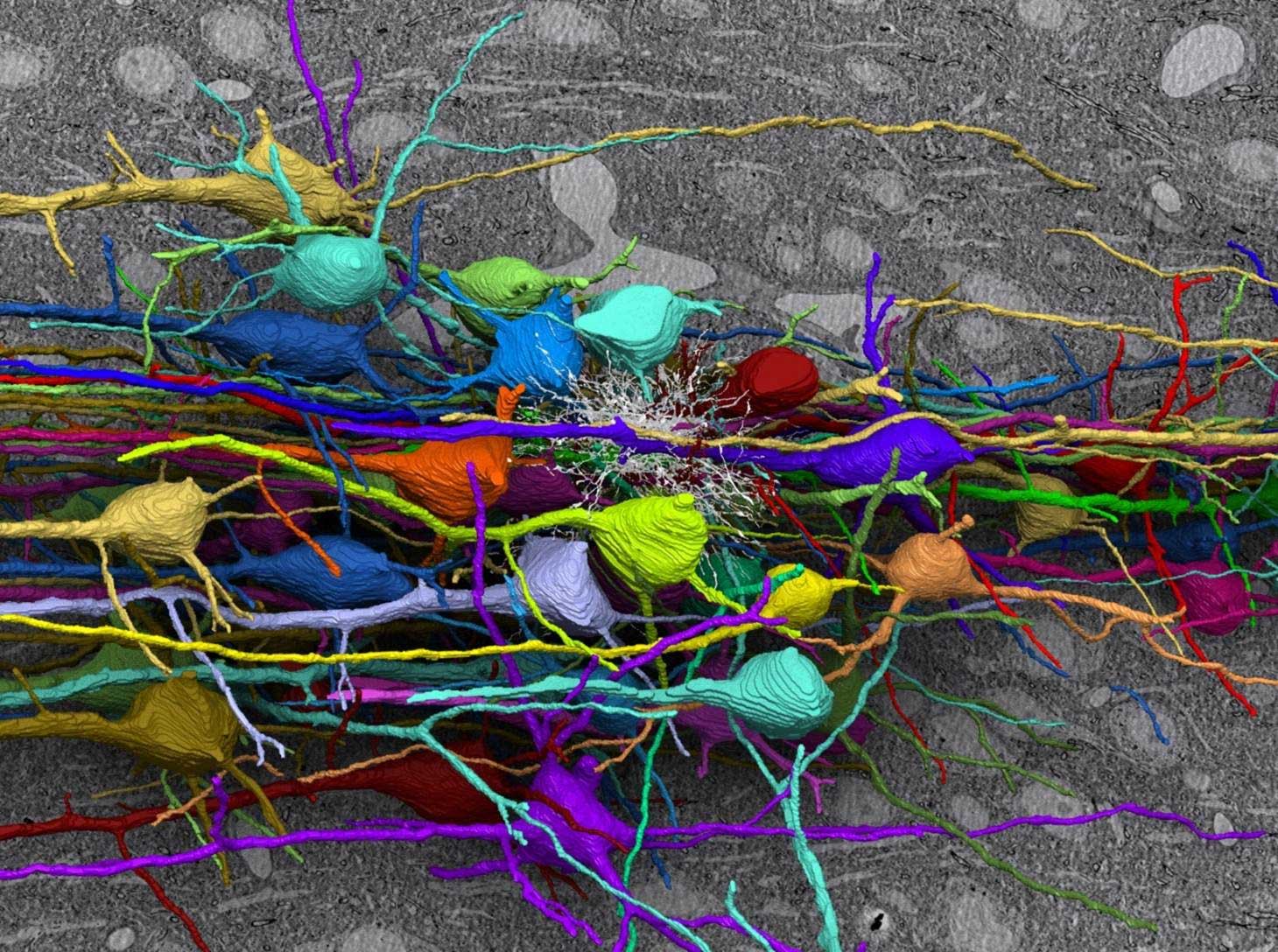
Connectomics: Unraveling the Wiring of Neural Networks
Working in connectomics means creating comprehensive maps of brain and nervous system networks. Your research includes the identification and measurement of all parts of each neuron: the soma, dendrites, axonal path and branching patterns and combining that data with the synapses and gap junctions of the entire circuit.
Your microscopy challenges are extensive; submicron resolution is required over long distances inside large volumes of dense and complicated tissues.
The Topological Lower Bound of Boltzmann Entropy: Resolving the Pure Top Boundary Condition through Proton Phase-Locking (v.01)
We establish a fundamental, non-zero lower bound for thermodynamic entropy by mapping Ludwig Boltzmann’s classical relation onto the rigid topological boundaries of GLAB chronal dynamics. In standard statistical mechanics, the number of microstates is treated as an abstract mathematical variable capable of reducing to unity , which phenomenologically implies an absolute zero entropy state . We demonstrate that this boundary condition is physically unattainable because the minimal, topologically closed space-phase cell possesses an irreversible internal structure dictated by the free proton configuration. Characterizing the stable proton as an asymmetric quantum “pure top” subject to the Janibekov instability, we prove that it inherently occupies a degenerate phase space composed of 2 intrinsic spin projections and 3 spatial rotational axes. This yields a strict, immutable minimum statistical weight of. Consequently, the absolute minimum entropy of any isolated domain in our universe is bounded by the Proton Constant:. We mathematically demonstrate that if this lower bound were violated, the phase-locking mechanism governing stellar nucleosynthesis would collapse, rendering the existence of periodic nuclear cycles and stable matter impossible.

Google releases new privacy controls for activity history, personalization
Google is rolling out new privacy controls for Search services and Google Play, giving you more control over saved history and personalized recommendations.
In an email titled “New privacy settings for Search services,” sent to users and seen by Bleeping Computer, Google said it is “updating our settings to give you even more control over saved history and personalized recommendations across Google Search services and Google Play.”
Google noted that Search services include “Search, Maps, Shopping, Hotels, Flights, Translate, and News,” and users will see the change in their Google Account in the next few days.
Google Is Mapping the Human Brain… and It Gets Terrifying
Google is using AI to map the human brain, generate synthetic neurons, and speed up one of the most ambitious neuroscience projects ever attempted. But as brain mapping, connectomics, and AI brain decoding move forward, a terrifying question appears: what happens to mental privacy when machines can understand the brain better than we do?
This mini-documentary explores Google’s brain mapping research, synthetic neurons, AI mind decoding, neural privacy, and the future of human thought in the age of artificial intelligence.
CHAPTERS:
00:00 Google’s Brain Mapping Project.
02:00 The Scale of the Human Brain.
04:36 Synthetic Neurons Explained.
06:40 AI Is Already Decoding Thoughts.
10:15 The Rise of Neural Privacy.
14:51 Brain Maps and the Future of AI
17:11 Who Owns Your Mind?
*******************
🌟 Reverse-Engineer ANY YouTube Channel in Seconds with the World’s First OS for Creators — OverseerOS:
https://www.overseeros.com/
*******************
Welcome to AI Uncovered, your ultimate destination for exploring the fascinating world of artificial intelligence! Our channel delves deep into the latest AI trends and technology, providing insights into cutting-edge AI tools, AI news, and breakthroughs in artificial general intelligence (AGI). We simplify complex concepts, making AI explained in a way that is accessible to everyone.
At AI Uncovered, we’re passionate about uncovering the most captivating stories in AI, including the marvels of ChatGPT and advancements by organizations like OpenAI. Our content spans a wide range of topics, from science news and AI innovations to in-depth discussions on the ethical implications of artificial intelligence. Our mission is to enlighten, inspire, and inform our audience about the rapidly evolving technology landscape.
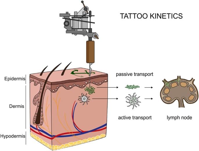
Synchrotron-based ν-XRF mapping and μ-FTIR microscopy enable to look into the fate and effects of tattoo pigments in human skin
Schreiver, I., Hesse, B., Seim, C. et al. Sci Rep 7, 11,395 (2017). https://doi.org/10.1038/s41598-017-11721-z.
Scientists Put a Fruit Fly’s Brain in a Computer Simulation… What It Did Is Now Scaring Scientists
Scientists have achieved an incredible breakthrough by recreating the brain of a fruit fly inside a computer simulation. By mapping around 140,000 neurons and millions of connections, they built a digital brain that can sense its environment, process information, and even control a virtual body. In the simulation, the digital fly was able to search for food, respond to stimuli, and show behaviors that were not directly programmed by scientists. This discovery shows how powerful neural connections are in generating behavior. It also raises fascinating questions about the nature of intelligence, consciousness, and whether complex brains—including ours—could one day be simulated in computers.
sources
https://eon.systems/updates/embodied-brain-emulation.
Research Paper for more information.
https://marginalrevolution.com/margin…
#Science.
#Neuroscience.
#ArtificialIntelligence.
#BrainSimulation.
#FruitFlyBrain.
#Connectome.
#FutureTech.
#ComputerSimulation.
#NeuralNetworks.
#ScienceDiscovery
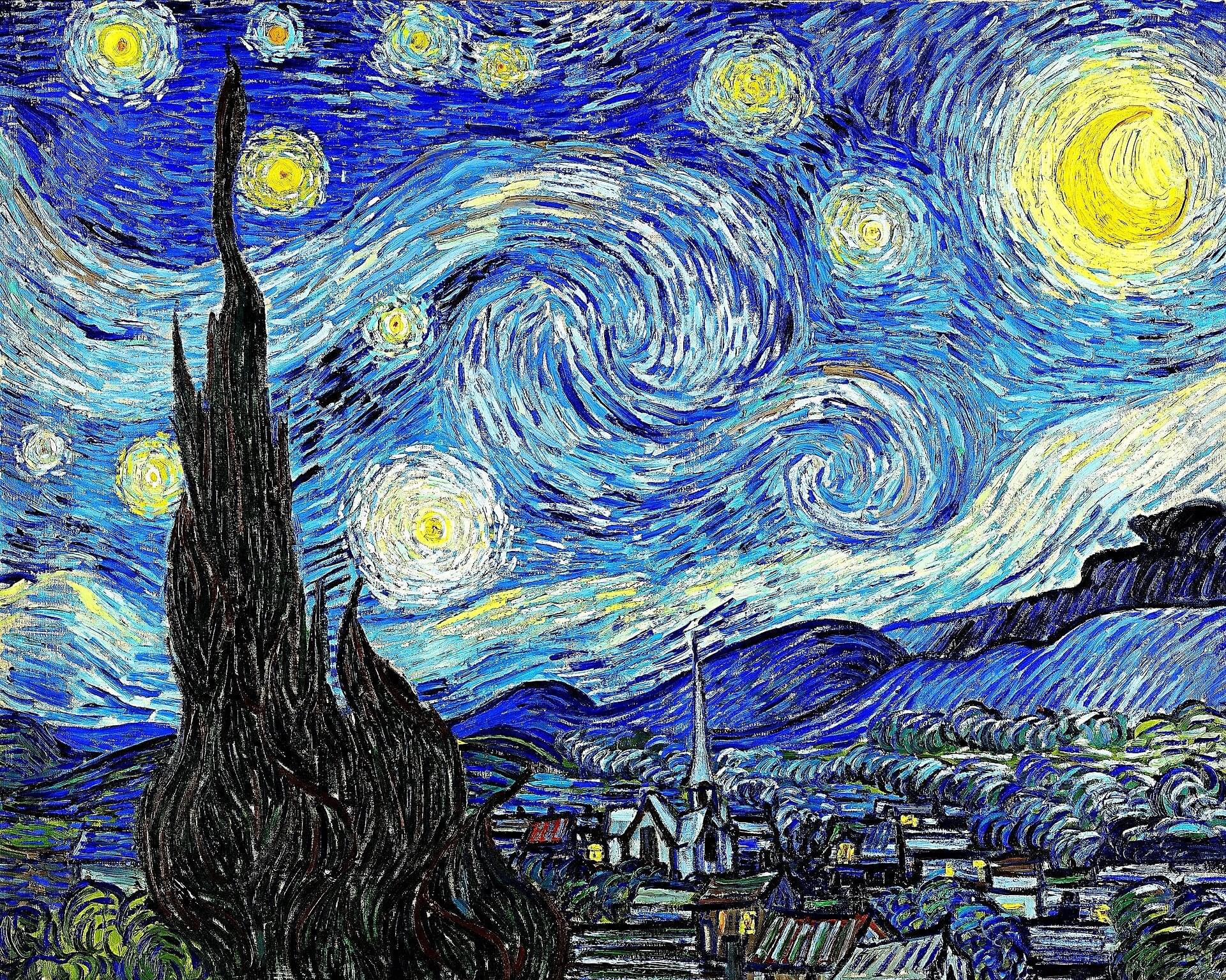
New art test could help museums spot fake Van Goghs without touching paintings
A new study published in the peer-reviewed journal Surface Topography: Metrology and Properties introduces a pioneering, noninvasive technique that can distinguish authentic artworks from forgeries, offering museums, collectors, and auction houses a major advantage in tackling art fraud.
The study, developed at the Université Polytechnique Hauts-de-France, introduces a method that analyzes the microscopic “texture” of a painting by converting high-resolution images into 3D-like maps, allowing researchers to measure how rough or detailed the surface is using fractal dimensions. This measurement captures subtle patterns created by an artist’s brushwork—patterns so consistent that they act like a morphological signature unique to that artist.
Using works attributed to Vincent van Gogh, the researchers showed that the method can reliably distinguish between authentic paintings and known forgeries. In tests, the well-documented fake “The Plowmen” was identified as a strong outlier, while the recently authenticated “Sunset at Montmajour” aligned closely with Van Gogh’s known works.