Researching are throwing lots of ideas at the wall to see what sticks, but the U.S. just put some serious cash behind the idea of 3D bioprinting.
Category: bioprinting
Programmable Macrophage Mimics for Inflammatory Meniscus Regeneration via Nanotherapy
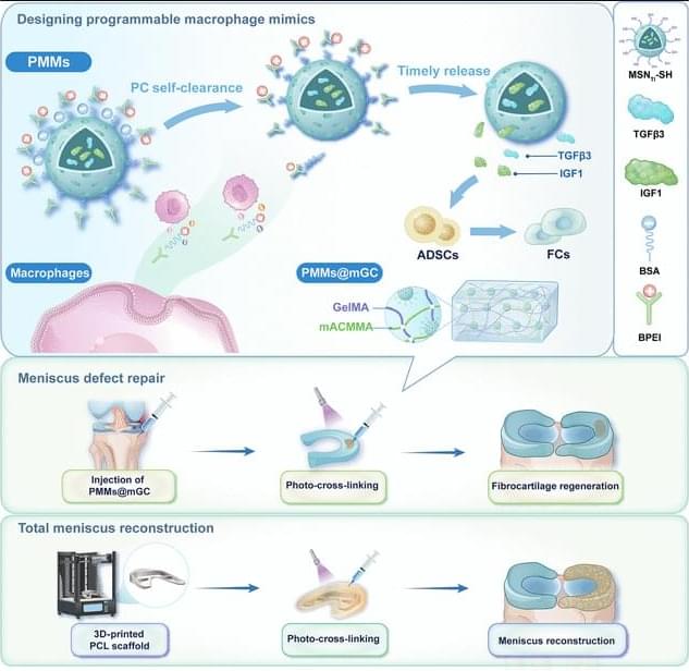
JUST PUBLISHED: programmable macrophage mimics for inflammatory meniscus regeneration via nanotherapy
Click here to read the latest free, Open Access Article from Research.
The meniscus is a fibrocartilaginous tissue and organ in the human knee joint that serves critical functions, including load transmission, shock absorption, joint stability, and lubrication. Meniscal injuries are among the most common knee injuries, typically caused by acute trauma or age-related degeneration [1– 3]. Minor meniscal injuries are usually treated with in situ arthroscopic procedures or conservative methods, whereas larger or more severe injuries often necessitate total meniscus replacement. Recent advances in materials science and manufacturing techniques have enabled transformative tissue-engineering strategies for meniscal therapy [4, 5]. Several stem cell types, including synovium-derived mesenchymal stem cells, bone-marrow-derived mesenchymal stem cells, and adipose-derived stem cells (ADSCs), have been investigated as candidate seed cells for meniscal regeneration and repair. Notably, ADSCs are clinically promising because of their ease of harvest, high inducibility, innate anti-inflammatory properties, and potential to promote fibrocartilage regeneration [6– 8]. Our group has developed a series of decellularized matrix scaffolds for auricular, nasal, tracheal, and articular cartilage repair using 3-dimensional (3D) bioprinting techniques, successfully repairing meniscus defects and restoring physiological function [9– 12]. However, current tissue-engineering strategies for meniscus defect repair commonly rely on a favorable regenerative microenvironment. Pathological conditions such as osteoarthritis (OA) [13 – 16], the most prevalent joint disorder, often create inflammatory environments that severely hinder meniscus regeneration [17 – 21]. Moreover, meniscal injury exacerbates the local inflammatory milieu, further impeding tissue healing and inevitably accelerating OA progression. Therefore, there is an urgent need to establish a cartilaginous immune microenvironment that first mitigates early-stage inflammation after meniscal injury and then sequentially promotes later-stage fibrocartilage regeneration [22 – 25].
Currently, targeted regulation using small-molecule drug injections is commonly employed to treat inflammatory conditions in sports medicine [26,27]. Most of these drugs exhibit broad-spectrum anti-inflammatory effects and inevitably cause varying degrees of side effects by activating nonspecific signaling pathways. Polyethyleneimine is a highly cationic polymer. It is widely used to modulate inflammation by adsorbing and removing negatively charged proinflammatory cytokines, including tumor necrosis factor-α (TNF-α), interleukin-1β (IL-1β), and interleukin-6 (IL-6), via electrostatic interactions [28–31]. Notably, modifying polyethyleneimine into its branched form (branched polyethyleneimine [BPEI]) has been shown to improve cytocompatibility and enhance in vivo metabolic cycling.
The Incredible Science of Bioprinting
Dive into the remarkable world of bioprinting in this comprehensive video. We’ll be exploring the core concepts of bioprinting — a pioneering technique that uses biological materials to create structures that mimic natural tissues, organs, and even cells. Understand the sophisticated science behind this process, and learn how bio-inks are formulated and layered to build live cells. We’ll also embark on a historical journey, tracing the origins and evolution of bioprinting, and how it is reshaping modern medicine. From overcoming organ shortages to paving the way for personalized treatments, bioprinting is revolutionizing healthcare. Join us as we unpack this fascinating technology and its promising future.
For more information on how patents can help drive your innovation.
visit https://www.patsnap.com/
From concept to commercialization, Patsnap’s innovation intelligence platform breaks boundaries to connect innovators with insights.
Follow us on social for more!
LinkedIn: / patsnap.
Facebook: / patsnap.
Twitter: / patsnap.
Instagram: / _patsnap.
PatSnap (UK) Limited (PatSnap) hereby disclaims all representations and warranties, express or implied, with respect to information, advertisements and all other ad-hoc materials published by us. PatSnap does not represent or warrant that the information accessible on this material is accurate, complete or current, and makes no claims or warranties of any kind, including the suitability of information, or merchantability and fitness for a particular purpose. It is the sole responsibility of the Recipient of such information to comply with all applicable laws, rules and regulations and they are responsible for meeting any regulatory, compliance and legal obligations. All due-diligence and vetting of services are the sole responsibility of the Recipient.
Droplet-based bioprinting Reviews Methods Primers
(DBB) enables the fabrication of three-dimensional structures from biomaterials. In this Primer, Gupta and colleagues describe the various DBB modalities and their applications, considerations for the selection of bioinks and substrates, methods to assess printability, the limitations of DBB and their solutions, and potential advances that might be brought about by incorporating new technologies.
A pill that prints bio-ink for damaged tissue repair
EPFL researchers have demonstrated the first pill-sized bioprinter that can be swallowed and guided within the gastrointestinal tract, where it directly deposits bio-ink over damaged tissues to support repair.
Soft tissue injuries of the gastrointestinal tract, like ulcers or hemorrhages, can currently be treated only with some form of surgery, which is invasive and may not result in permanent repair. Bioprinting is emerging as an effective treatment that deposits biocompatible “ink”—often made of natural polymers derived from seaweed—directly over the site of tissue damage, creating a scaffold for new cell growth. But like traditional surgical tools, these kinds of bioprinters tend to be bulky and require anesthesia.
At the same time, “untethered” technologies are being developed to perform medical interventions without a physical connection to external equipment. For example, ingestible “smart capsules” can be guided to drug delivery sites using external magnets. But these devices are designed to travel through liquids, and their movements become unpredictable when they touch the tissue wall.
Breakthrough 3D Bioprinted Mini Placentas May Help Solve “One Of Medicine’s Great Mysteries”
To address these shortcomings, the team behind the latest study turned to bioprinting – a type of 3D printing that uses living cells and cell-friendly materials to create 3D structures. They took trophoblast cells and mixed them with a synthetic gel before 3D-printing them in precise droplets.
The printed cells then grew into miniature placentas, and the researchers compared them to organoids made via traditional manual methods.
“The organoids we grew in the bioprinted gel developed differently to those grown in an animal-derived gel, and formed different numbers of trophoblast sub-types. This highlighted that the environment organoids are grown in can control how they mature,” first author Dr Claire Richards said.
Adaptable 3D bioprinting technique can boost engineered tissue output
The field of tissue engineering aims to replicate the structure and function of real biological tissues. This engineered tissue has potential applications in disease modeling, drug discovery, and implantable grafts.
AI turns printer into a partner in tissue engineering
In 3D bioprinting, researchers use living cells to create functional tissues and organs. Instead of printing with plastic, they print with living cells. This comes with great challenges. Cells are fragile and wouldn’t survive a regular 3D printing process. That’s why Levato’s team developed a special bio-ink, a mix of living cells and nourishing gels that protect the cells during the printing process.
With the advancements in bio-inks, layer-by-layer 3D bioprinting became possible. But this method is still time-consuming and puts a lot of stress on the cells. Researchers from Utrecht came up with a solution: volumetric bioprinting.
Volumetric bioprinting is faster and gentler on cells. Using cell-friendly laser light, a 3D structure is created all at once. “To build a structure, we project a series of light patterns into a spinning tube filled with light-sensitive gel and cells,” Levato explains. “Where the light beams converge, the material solidifies. This creates a full 3D object in one go, without having to touch the cells.” To do this, it is crucial to know exactly where the cells are in the gel. GRACE now makes that possible.
Advanced model unlocks granular hydrogel mechanics for biomedical applications
Researchers at the University of Illinois Urbana-Champaign have developed a novel framework for understanding and controlling the flow behavior of granular hydrogels—a class of material made up of densely packed, microscopic gel particles with promising applications in medicine, 3D bioprinting, and tissue repair.
The new study, published in Advanced Materials, was led by chemical and biomolecular engineering professors Brendan A. Harley and Simon A. Rogers, whose research groups specialize in biomaterials engineering and rheology, respectively.
Granular hydrogels have a unique ability to mimic the mechanical properties of living tissue, which makes them ideal candidates for encapsulating and delivering cells directly into the body. By integrating material synthesis and characterization with rheological modeling, the researchers created a predictive model that captures the essential physics of how granular hydrogels deform—reducing a complex problem to a few controllable parameters.