Toggle light / dark theme

Does a prospective father’s gut microbiota matter?

Germline cells play a key role in the transmission of phenotypes and physiological adaptations to subsequent generations (1). Over a century ago, August Weismann proposed that changes in somatic cells cannot be passed on to germ cells or offspring, a theory known as the Weismann barrier (2). Nevertheless, recent studies have proven that the Weismann barrier is permeable, and information can pass from soma to germline and modulate offspring phenotypes. In the past decade, there has been tremendous interest and progress in understanding how an altered microbiome (dysbiosis) affects different somatic cells that compose body tissues, such as brain, liver, heart, kidney, and lungs (3). Nevertheless, whether gut microbiome dysbiosis can exert an influence on the mammalian germline cells (i.e., gut to germline), and ultimately nonexposed offspring, remains unclear.

To tackle this research question, my colleagues and I established an inducible model of gut microbiota dysbiosis in isogenic male mice, using ad lib nonabsorbable antibiotics (nABX) that cannot cross the epithelial barrier of the gut (4). As expected, 6 weeks of low-dose nABX treatment led to a physiologically significant dysbiosis, which is reversible and gradually normalized to a physiologically healthy gut microbiota after 8 weeks of nABX withdrawal (6 weeks + 8 recovery). The induced dysbiosis after 6 weeks of nABX had no appreciable effects on male body weight, growth, or fertility. No nABx residues were detected in the serum or testes of treated males, which confirmed that any distal tissue responses are gut dysbiosis–induced rather than systemic drug effects.

We then examined physiological changes in the male reproductive system in response to 6 weeks of dysbiosis. Dysbiotic males had smaller testes, lower sperm count, and more abnormally shaped sperm. Histological analysis uncovered a wide range of anatomical abnormalities in testes of dysbiotic males, including increased number of abnormal seminiferous tubules, reduced epithelial thickness, and absence of mitotic compartments, which were not observed in control testes. Testicular metabolomic profiles revealed that testes clustered according to gut microbiota status and exhibited dysregulated sphingolipids, glycerophospholipids, and endocannabinoids, all known to play pivotal roles in germ cell function. Moreover, in dysbiotic male testes, spermatogenesis-regulating genes were misexpressed—most notably leptin, a reproductive hormone, was strongly down-regulated.

Babies’ poor vision may help organize visual brain pathways

Incoming information from the retina is channeled into two pathways in the brain’s visual system: one that’s responsible for processing color and fine spatial detail, and another that’s involved in spatial localization and detecting high temporal frequencies. A new study from MIT provides an account for how these two pathways may be shaped by developmental factors.

Newborns typically have poor visual acuity and poor vision because their retinal cone cells are not well-developed at birth. This means that early in life, they are seeing blurry, color-reduced imagery. The MIT team proposes that such blurry, color-limited vision may result in some specializing in low spatial frequencies and low color tuning, corresponding to the so-called magnocellular system. Later, with improved vision, cells may tune to finer details and richer color, consistent with the other pathway, known as the parvocellular system.

To test their hypothesis, the researchers trained computational models of vision on a trajectory of input similar to what human babies receive early in life—low-quality images early on, followed by full-color, sharper images later. They found that these models developed processing units with receptive fields exhibiting some similarity to the division of magnocellular and parvocellular pathways in the human visual system. Vision models trained on only high-quality images did not develop such distinct characteristics.

New neurons continue to form in the adult human hippocampus: Study

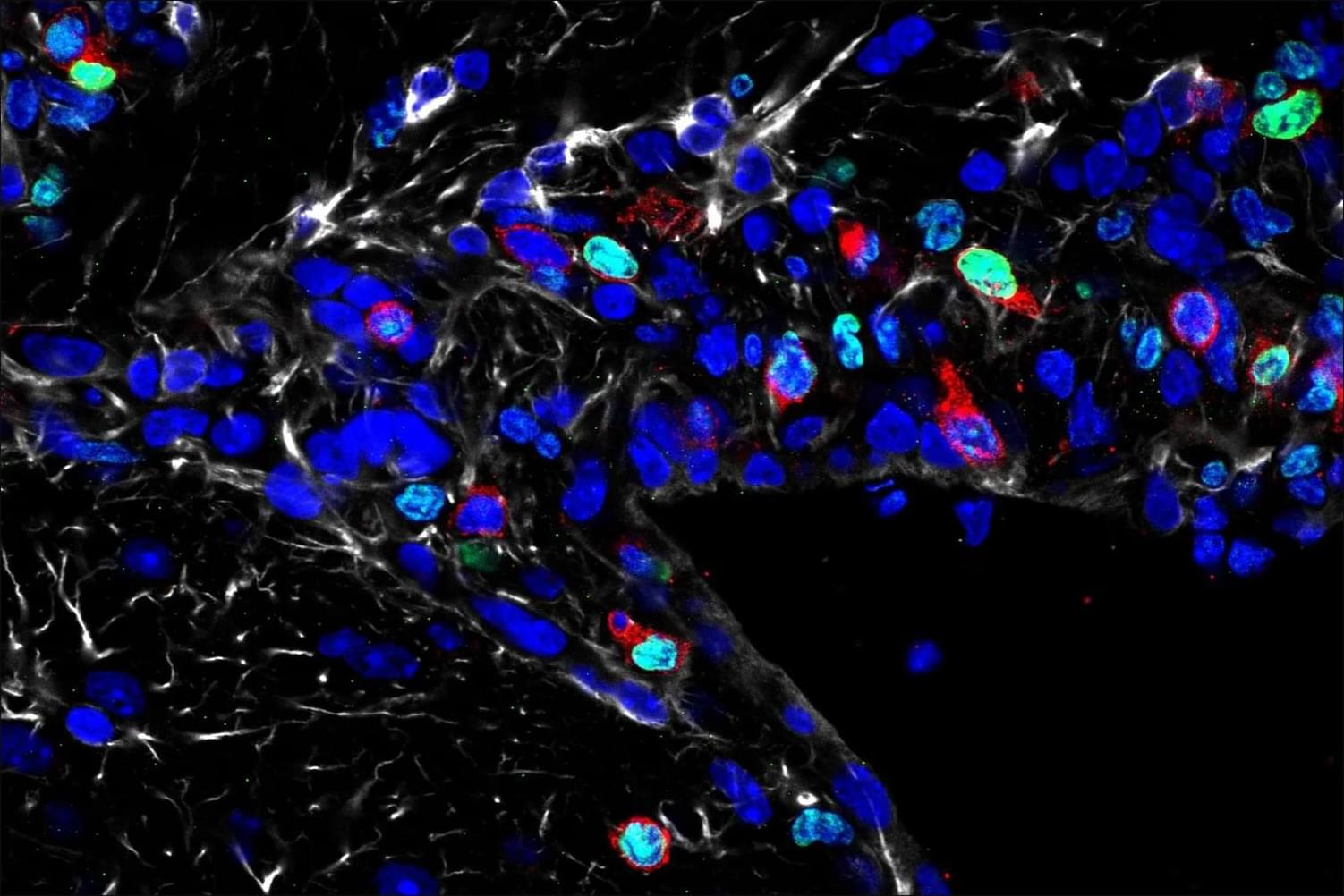
A study in the journal Science presents compelling new evidence that neurons in the brain’s memory center, the hippocampus, continue to form well into late adulthood. The research from Karolinska Institutet in Sweden provides answers to a fundamental and long-debated question about the human brain’s adaptability.

The hippocampus is a brain region that is essential for learning and memory and involved in emotion regulation. Back in 2013, Jonas Frisén’s research group at Karolinska Institutet showed in a high-profile study that can form in the hippocampus of adult humans. The researchers then measured carbon-14 levels in DNA from , which made it possible to determine when the cells were formed.

“How to build conscious machines” by Michael Timothy Bennett

This is a ~1 hour talk by Michael Timothy Bennett (https://michaeltimothybennett.com/) on his ideas around computation and consciousness.

Direct link to the thesis preprint: https://osf.io/preprints/thesiscommons/wehmg_v1?view_only=

YT: https://www.youtube.com/@michaeltimothybennett.
X: https://twitter.com/MiTiBennett

Neuropsychological evidence of a third visual pathway specialized for social perception

Recent evidence suggests the existence of a neural pathway specialized for social perception projecting between the well-established “what” and “where” pathways. A new study of neuropsychological patients demonstrates that this social pathway is causally essential for recognizing dynamic facial expressions.

Researchers develop two-layer neural model that matches complex visual processing in the brain

Neuroscientists want to understand how individual neurons encode information that allows us to distinguish objects, like telling a leaf apart from a rock. But they have struggled to build computational models that are simple enough to allow them to understand what individual neurons are doing.

To address this challenge, researchers in the Stringer and Pachitariu labs at Janelia set out to create a simpler model to explain what’s going on in the —the first stop in the brain for . Their paper is published in the journal Nature Communications.

“We are trying to build a model that can predict the visual responses of each individual neuron,” says Fengtong Du, a graduate student in the Stringer Lab who led the new research.