The US Endangered Species Act compels the government to identify species at risk of extinction and devise plans to restore populations and the habitats they depend on. It has seen some spectacular successes, such as the restoration of the bald eagle to much of its original range. But over 2,300 plant and animal populations remain on the list, requiring ongoing government intervention.
On Thursday, it was announced that all of those species would see their genomes sequenced and tissue samples preserved to aid future conservation efforts. The work will be done by a partnership between two unexpected parties. One is the US government, which has generally attempted to undercut the Endangered Species Act as part of its anti-regulatory efforts. It is joined by Colossal Biosciences, a biotech company that has a controversial take on what actually constitutes a species.
Colossal has always said it had a conservation focus, but its headline-grabbing efforts have been directed toward restoring species that have been driven to extinction. It intends to do that by developing a combination of gene editing and reproductive technologies that it expects it can profitably license. But its dire wolf announcement, in which only a tiny handful of genetic changes were edited in to grey wolves, have raised some questions about its seriousness regarding these efforts.
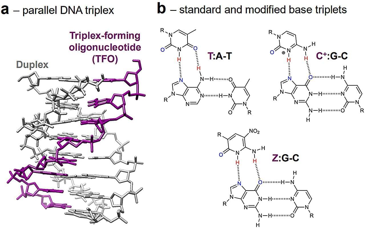
A new method for recognizing and targeting DNA that dramatically expands the range of genetic sequences scientists can identify has been developed by experts at the University of Portsmouth. Published this week in Nature Communications, the research opens new possibilities for gene-targeting technologies, molecular diagnostics and DNA nanotechnology.
Dr. David Rusling, associate professor in bioengineering from the University of Portsmouth’s School of Medicine, Pharmacy and Biomedical Sciences, said, Our lab develops synthetic molecules that can recognize and bind to unique gene sequences. By introducing synthetic DNA bases into these molecules, we’ve been able to significantly improve how they recognize their targets.
I’ve worked in this area for around 20 years, and this is the first time we’ve had a system that combines strong recognition under physiological conditions with building blocks that are commercially available to other researchers.
Discover the groundbreaking world of lab-grown organs in our latest YouTube Shorts! In “Lab-Grown Organs: Revolutionizing Transplants,” we explore how scientists are utilizing bioprinting, scaffold tissue engineering, and induced pluripotent stem cells to create functional organs like kidneys, livers, and hearts. This innovative approach not only eliminates transplant waiting lists but also uses a patient’s own cells, reducing the risk of rejection. Join us as we unveil the future of organ transplantation and the incredible advancements in organogenesis!
If you find this video enlightening, don’t forget to like and share it with your friends!
Michael Levin is a developmental and synthetic biologist at Tufts University whose work sits at the intersection of biology, bioelectricity, artificial life, regenerative medicine, synthetic biology, computer science, cognitive science, and philosophy of mind. He is known for his research on how cells communicate, make decisions, build bodies, repair tissues, and form collective intelligence through bioelectric signals. His work on Xenobots and Anthrobots has opened new questions about living robots, synthetic life forms, biological machines, morphogenesis, basal cognition, cellular intelligence, regeneration, cancer, aging, and the nature of mind beyond the brain.
In this conversation, Michael Levin and I explore whether mind and intelligence are binary or exist on a continuum, why cognition may be much older than brains, and how systems from cells to humans can pursue goals in different ways. We discuss the TAME framework, the spectrum of persuadability, cognitive light cones, bioelectricity, gap junctions, multicellular intelligence, Xenobots, Anthrobots, kinematic self-replication, neural wound healing, emergence, physicalism, mathematics, Platonic space, algorithms, bubble sort, Turing machines, evolution, human creativity, artificial intelligence, regenerative medicine, and the future of biology. This episode is for anyone interested in philosophy, consciousness, mind, intelligence, synthetic biology, developmental biology, AI, complex systems, evolution, and the deeper question of what it means for matter to become alive, intelligent, or aware.
If you enjoyed the episode, please consider leaving a like, subscribing, and leaving a review on Youtube, Spotify and Apple. #philosophy #science.
Socials: Spotify: https://open.spotify.com/show/46hnFSg… Podcasts: https://podcasts.apple.com/us/podcast… Linkedin: / masud-gaziyev Instagram (public): / philosophy.everyday Instagram (private): / masud.gaziyev Support the work: https://buymeacoffee.com/philosophy.e… Get new episodes, guest announcements, reading notes, and ideas worth thinking about. Subscribe here: https://philosophyeveryday.beehiiv.com/ Chapters: 00:00 Mind Beyond the Brain 01:19 Is Mind Older Than the Brain? 04:06 Why Intelligence Is Not All-or-Nothing 06:58 How to Interact With Different Kinds of Minds 09:54 From Single Cells to Collective Intelligence 13:17 How Cells Build Bigger Goals 16:05 Life Recreated — Xenobots and Anthrobots 18:54 Where Do New Behaviours Come From? 21:57 Synthetic Life and the Limits of Evolution 35:01 What Happens When Biology Is Freed? 43:00 Why Biology Eventually Leads to Mathematics 46:07 Is “Emergence” Just a Fancy Word for Surprise? 53:11 Platonic Space: A Strange New Map of Reality 01:03:21 What We Received from Platonic Space 01:11:24 Human Evolution, Technology, and the Patterns Behind Progress 01:16:43 Regeneration, Cancer, and Aging. Apple Podcasts: https://podcasts.apple.com/us/podcast… Linkedin: / masud-gaziyev. Instagram (public): / philosophy.everyday. Instagram (private): / masud.gaziyev. Support the work: https://buymeacoffee.com/philosophy.e…
Get new episodes, guest announcements, reading notes, and ideas worth thinking about.
In 2012, I sat down with Dr. James Hughes, bioethicist, sociologist, and executive director of the Institute for Ethics and Emerging Technologies.
Fourteen years later, the questions we wrestled with have only sharpened.
Why are transhumanist atheists so often drawn to Buddhism? Is optimism rational, or just a posture we adopt to keep moving? What does it mean to redesign the human being, and which democratic institutions are ready to respond when we do?
James does not flinch from any of it. He talks about his first book Citizen Cyborg, the then forthcoming Cyborg Buddha, moral enhancement, animal uplift, and what our actual chances are of surviving the technological singularity.
What struck me most was his refusal to retreat into easy camps.
Not a cheerleader, not a doomsayer. Someone who interrogates the world and engages it on its own terms.
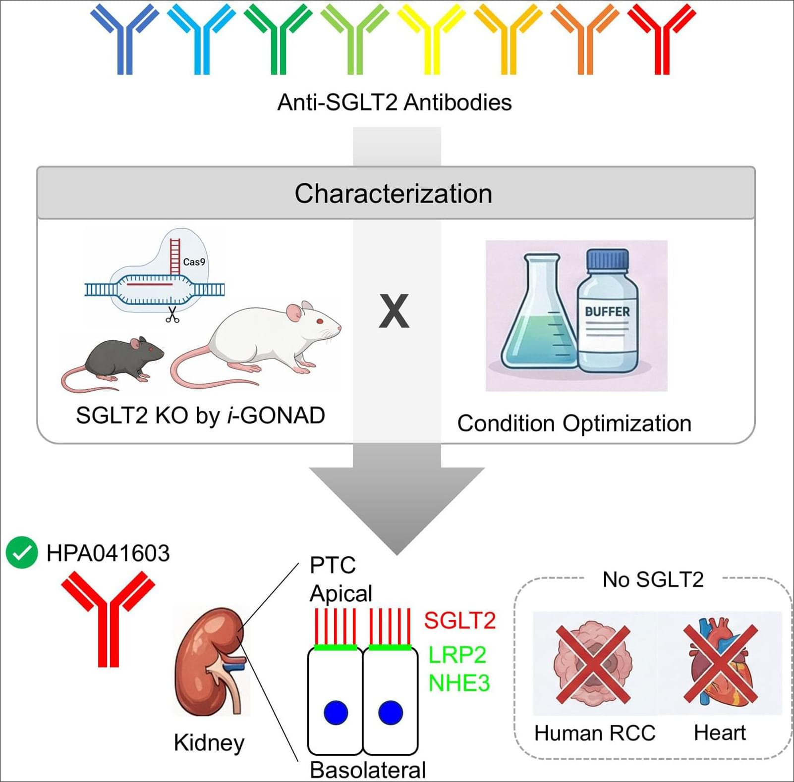
BACKGROUND: SGLT2 (sodium-glucose cotransporter 2) mediates renal glucose reabsorption, and its pharmacological inhibition exerts cardio-and renoprotective benefits. Despite widespread clinical interest, reliable detection of SGLT2 protein remains challenging due to concerns regarding antibody specificity. METHODS: Eight commercially available anti-SGLT2 antibodies were evaluated by immunohistochemistry and Western blotting using kidneys and hearts from genetically engineered Sglt2-deficient mice and rats. Human kidney tissues, including renal cell carcinoma samples, were also examined. RESULTS: Among the antibodies tested, ab306558 and HPA041603 showed specific immunostaining in rodent kidneys, with minimal background in wild-type tissues and complete absence of staining in Sglt2-deficient samples. However, ab306558 was unsuitable for human samples because of nonspecific staining.
What will the world really look like in 100 years?
Forget flying cars, impossible megacities, and science-fiction fantasies. This documentary explores a realistic vision of life in the year 2,126 based on current trends in artificial intelligence, climate adaptation, biotechnology, energy, space exploration, economics, and human evolution.
How will cities change as the planet warms? What happens when AI becomes part of everyday life? Will humans live to 120 years? Will neural implants blur the line between biology and technology? Could Mars become a permanent home for thousands of people? And what happens to society when work, truth, privacy, and even human identity are redefined?
Travel one century into the future and discover a world that is both familiar and radically different from our own. A world shaped by the choices humanity is making right now.
From climate-engineered cities and fusion-powered civilizations to Martian settlements, artificial intelligence, genetic medicine, digital consciousness, and the search for life beyond Earth, this is a deep exploration of the most plausible future awaiting our species.
We’re launching a new series at StayCurious Metabolism called Peptides Plus, where we’ll explore the most promising tools available today—and the innovations that may shape tomorrow. We have dozens of deep dives planned, covering everything from emerging therapeutics to cutting-edge performance and longevity interventions.
Chapters. 0:00 — Superhuman Biology Is Already Starting. 2:40 — Beyond GLP-1: Fat Loss Without Muscle Loss. 7:28 — Gene Editing, CRISPR, and the Future of Disease Cure. 14:57 — Cellular Reprogramming and Biological Age Reset. 18:49 — MicroRNAs, Mitochondria, and What Comes Next.
DNA is composed of long chains that act as the blueprint for living organisms. In genetic engineering, scientists cut DNA at specific sites and join the resulting fragments to other DNA sequences, enabling applications such as advanced crop breeding, treatment of genetic diseases, and the generation of animal models for drug discovery.
Assembling short DNA fragments requires overhanging sequences, known as sticky ends, to facilitate efficient binding. However, generating sticky ends requires precise cutting at targeted sites, which remains challenging with current technologies.
A Japanese research group has developed a silver nanoparticle-based technology to precisely cut and join DNA at targeted sites, achieving two to five times higher DNA assembly efficiency than conventional restriction enzyme methods. These findings were published in the journal Nucleic Acids Research.