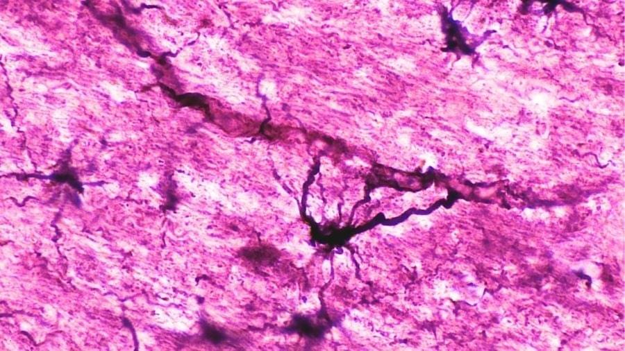
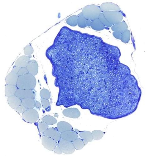
The cell membrane plays a critical role in regulating substance exchange, signal transduction, and energy conversion, making it essential for maintaining homeostasis and responding to environmental stimuli.
A robot butler sounds like a nice idea, but the technology has its drawbacks.
Johns Hopkins scientists say they have used 3D imaging, special microscopes and artificial intelligence (AI) programs to construct new maps of mouse brains showing a precise location of more than 10 million cells called oligodendrocytes. These cells form myelin, a protective sleeve around nerve cell axons, which speeds transmission of electrical signals and support brain health.
Published online Feb. 18 in Cell and funded by the National Institutes of Health, the maps not only paint a whole-brain picture of how myelin content varies between brain circuits, but also provide insights into how the loss of such cells impacts human diseases such as multiple sclerosis, Alzheimer’s disease and other disorders that affect learning, memory, sensory ability and movement, say the researchers. Although mouse and human brains are not the same, they share many characteristics and most biological processes.
“Our study identifies not only the location of oligodendrocytes in the brain, but also integrates information about gene expression and the structural features of neurons,” says Dwight Bergles, Ph.D., the Diana Sylvestre and Charles Homcy Professor in the Department of Neuroscience at the Johns Hopkins University School of Medicine. “It’s like mapping the location of all the trees in a forest, but also adding information about soil quality, weather and geology to understand the forest ecosystem.”
Picture a star-shaped cell in the brain, stretching its spindly arms out to cradle the neurons around it. That’s an astrocyte, and for a long time, scientists thought its job was caretaking the brain, gluing together neurons, and maintaining neural circuits. But now, a new study reveals that these supposed support cells that are spread all over the brain are as important as neurons in fear memory.
“Astrocytes are interwoven among neurons in the brain, and it seemed unlikely they were there just for housekeeping. We wanted to understand what they’re actually doing—and how they’re shaping neural activity in the process,” said Lindsay Halladay, assistant professor at the University of Arizona Department of Neuroscience and one of the study’s senior authors.
Halladay’s lab collaborated with researchers from the National Institutes of Health for this multi-institutional study, led by Andrew Holmes and Olena Bukalo of the Laboratory of Behavioral and Genomic Neuroscience.
Five years of ublituximab treatment in MultipleSclerosis demonstrated sustained reduction in relapse rates and confirmed disability progression, with safety profile consistent over time.
Question What is the long-term clinical efficacy and safety of ublituximab in people with relapsing multiple sclerosis (RMS)?
Findings In this trial including 985 adults, participants treated with continuous ublituximab for up to 5 years in the open-label extension study after completion of the randomized Study to Assess the Efficacy and Safety of Ublituximab in Participants With Relapsing Forms of Multiple Sclerosis (ULTIMATE) had significantly lower annualized relapse rate and confirmed disability progression than those initially treated with teriflunomide. The overall safety profile of ublituximab remained consistent with no new safety signals emerging with prolonged treatment.
Meaning Results suggest that early initiation of ublituximab and continued treatment over a period of 5 years provided sustained clinical benefits in participants with RMS.
🧠 AI in pathology cannot succeed without pathologists. As computational pathology advances, clinical expertise remains the critical link between algorithms and real-world impact.
In this discussion, Diana Montezuma, Pathologist and Head of R&D at IMP Diagnostics, explains why pathologist involvement is essential to building AI tools that are usable, clinically relevant, and truly valuable in practice.
👉 Read the discussion:
Pathologists play a key role in AI development for pathology – providing the expertise needed to bridge data and clinical application. To discuss this role and its importance in the development of computational pathology tools, we connected with Diana Montezuma, Pathologist and Head of the R&D Unit at IMP Diagnostics.
From your perspective, what is the most important contribution that diagnosticians bring to AI and algorithm development?
Pathologists bring essential clinical expertise and practical insight to any computational pathology project. Without their involvement, such initiatives risk becoming disconnected from real-world practice and ultimately failing to deliver meaningful clinical value.
The tiny fatty capsules that deliver COVID-19 mRNA vaccines into billions of arms may work better when they’re a little disorganized. That’s the surprising finding from researchers who developed a new way to examine these drug-delivery vehicles one particle at a time—revealing that cramming in more medicine doesn’t always mean better results.
The research was presented at the 70th Biophysical Society Annual Meeting, held in San Francisco from February 21–25, 2026.
Lipid nanoparticles, or LNPs, are microscopic bubbles of fat that can ferry fragile RNA molecules into cells. They were crucial to the success of mRNA vaccines, and scientists are now working to use them to deliver treatments for cancer, genetic diseases, and other conditions. But there’s a problem: only about 1% to 5% of the cargo inside LNPs actually gets released inside cells.
Teeth are composites of mineral and protein, with a bulk of bony dentin that is highly porous. This structure allows teeth to be both strong and sensitive. Besides calcium and phosphate, teeth contain trace elements such as zinc. Using complementary microscopy imaging techniques, a team from Charité Berlin, TU Berlin and HZB has quantified the distribution of natural zinc along and across teeth in 3 dimensions. The team found that, as porosity in dentin increases towards the pulp, zinc concentration increases 5~10 fold. These results help to understand the influence of widely-used zinc-containing biomaterials (e.g. filling) and could inspire improvements in dental medicine.
The paper is published in the journal VIEW.
Teeth have a complex structure: the dental pulp with the nerves is surrounded by dentin, a porous bony material, covered externally by enamel in the mouth and cementum in the roots. Although dentin is criss-crossed by countless micrometer-sized dentin tubules, teeth can withstand decades of cyclic, repeated forces. The density of the dentinal tubules increases towards the pulp, meaning that the dentin becomes increasingly porous towards the inside.
A new study demonstrates that computer models of gut metabolism can predict which probiotics will successfully establish themselves in a person’s gut and how different prebiotics affect production of health-promoting short-chain fatty acids. The findings are published in PLOS Biology by Sean Gibbons of the Institute for Systems Biology, US, and colleagues.
Probiotic and prebiotic supplements show highly variable results across individuals, making it difficult to predict who will benefit from these interventions. This variability comes from complex interactions between introduced probiotics, each person’s existing gut microbiota, and their diet.
In the new work, researchers first tested a metabolic model on data from two previous studies in which participants diagnosed with type 2 diabetes were given a placebo or probiotic/prebiotic mixture designed to improve glucose control and healthy participants were given a placebo or a probiotic treatment designed to treat recurrent Clostridioides difficile infections, respectively.
Ion channels are narrow passageways that play a pivotal role in many biological processes. To model how ions move through these tight spaces, pores need to be fabricated at very small length scales. The narrowest regions of ion channels can be just a few angstroms wide, about the size of individual atoms, making reproducible and precise fabrication a major challenge in modern nanotechnology.
In a study published in Nature Communications, researchers at The University of Osaka have addressed this challenge by using a miniature electrochemical reactor to create ultra-small pores approaching subnanometer dimensions.
In biological cells, ions flow in and out through channels in cell membranes. This ion flow is the basis for generating electrical signals, such as nerve impulses that trigger muscle contraction. The channels themselves are made of proteins and can have angstrom-wide narrow regions. Conformational changes of these proteins in response to external stimuli open and close the channels.