The brain of an octopus has some similarities to humans, and shows many signs of high intelligence.
Category: neuroscience – Page 672
Newton Howard (MIT Synthetic Intelligence Lab)– The Future of Brain Implants
Newton Howard, Director of the MIT Synthetic Intelligence Lab: http://www.techsylvania.co/speakers/newton-howard/
Check out the presentation on Slideshare: https://www.slideshare.net/techsylvania/newton-howard-mit
Brain Implants are Here: Blackrock’s Neuroport & Synchron’s Stentrode
Neurotechnology and Brain-Computer Interfaces are advancing at a rapid pace and may soon be a life-changing technology for those with limited mobility and/or paralysis. There are already two brain implants, Blackrock Neurotech’s NeuroPort and Synchron’s Stentrode, that have been approved to start clinical trials under an Investigational Device Exemption. In this video, we compare these devices on the merits of safety, device specifications, and capability.
Thanks to Blackrock Neurotech for sponsoring this video. The opinions expressed in this video are that of The BCI Guys and should be taken as such.
——–ABOUT US:——-
Harrison and Colin (The BCI Guys) are neurotech researchers and entrepreneurs dedicated to creating a brain-controlled future! Neurotechnology and brain-computer interfaces are devices that allow users to control machines with their thoughts and interact with technology in new ways. This revolutionary technology will change life as we know it, and soon will be as common as the touchscreen on your smartphone. Join us in learning about the brain-controlled future!
Support us: https://www.bciguys.com/support.
Follow us on Social Media!
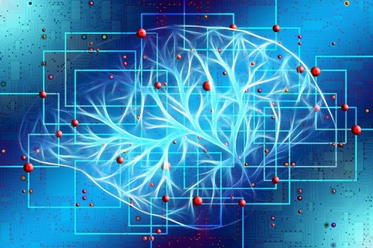
The Biological Basis of Network Control Theory in Brain Dynamics
Summary: Researchers have identified a correlation between control energy consumption and glucose metabolism in temporal lobe epilepsy. The mechanism provides a biological basis for the application of network control theory in the study of brain dynamics.
Source: USTC
A team led by researcher He Xiaosong from the University of Science and Technology (USTC) revealed the correlation between control energy consumption and glucose metabolism in temporal lobe epilepsy (TLE), providing the biological basis for the application of network control theory (NCT) in the study of brain dynamics.
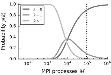
Extremely Scalable Spiking Neuronal Network Simulation Code: From Laptops to Exascale Computers
Year 2018 😗
State-of-the-art software tools for neuronal network simulations scale to the largest computing systems available today and enable investigations of large-scale networks of up to 10% of the human cortex at a resolution of individual neurons and synapses. Due to an upper limit on the number of incoming connections of a single neuron, network connectivity becomes extremely sparse at this scale. To manage computational costs, simulation software ultimately targeting the brain scale needs to fully exploit this sparsity. Here we present a two-tier connection infrastructure and a framework for directed communication among compute nodes accounting for the sparsity of brain-scale networks. We demonstrate the feasibility of this approach by implementing the technology in the NEST simulation code and we investigate its performance in different scaling scenarios of typical network simulations. Our results show that the new data structures and communication scheme prepare the simulation kernel for post-petascale high-performance computing facilities without sacrificing performance in smaller systems.
Modern neuroscience has established numerical simulation as a third pillar supporting the investigation of the dynamics and function of neuronal networks, next to experimental and theoretical approaches. Simulation software reflects the diversity of modern neuroscientific research with tools ranging from the molecular scale to investigate processes at individual synapses (Wils and De Schutter, 2009) to whole-brain simulations at the population level that can be directly related to clinical measures (Sanz Leon et al., 2013). Most neuronal network simulation software, however, is based on the hypothesis that the main processes of brain function can be captured at the level of individual nerve cells and their interactions through electrical pulses. Since these pulses show little variation in shape, it is generally believed that they convey information only through their timing or rate of occurrence.
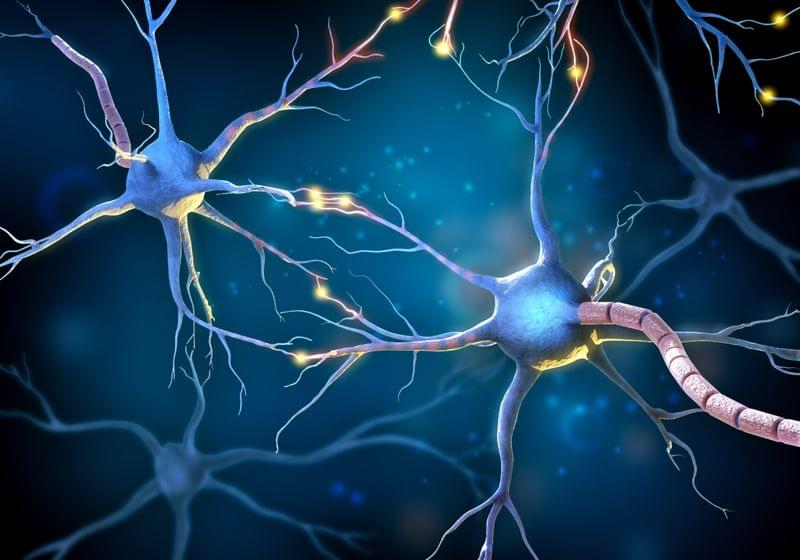
I got a chip implanted in a biohacking garage
In the underground movement known as, people are taking their health into their own hands. Biohacking ranges from people making simple lifestyle changes to extreme body modifications.
One popular form of focuses on nutrigenomics, where biohackers study how the foods they eat affect their genes over time. They believe they can map and track the way their diet affects genetic function. They use dietary restrictions and blood tests, while tracking their moods, energy levels, behaviors, and cognitive abilities.
Then there are grinders, a subculture of A grinder believes there’s a hack for every part of the body. Rather than attempting to modify our existing biology, grinders seek to enhance it with implanted technology.
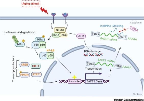
Unmasking BACE1 in aging and age-related diseases
The BACE1 enzyme has a rate-limiting role in the amyloidogenic pathway (see Glossary) and has been extensively studied for its neuronal functions[1]. Since 2000, intensive efforts have focused on developing small-molecule BACE1 inhibitors to reduce amyloid β (Aβ) production in Alzheimer’s disease (AD) brains. However, human clinical trials involving most BACE1 inhibitors were stopped at Phase 2/3 due to limited therapeutic benefits[2]. BACE1 inhibitors act by reducing Aβ-related pathologies in AD brains, that is, they are used to treat the symptoms rather than the underlying disease.
