Long-term melatonin use may secretly strain the heart—raising the risk of failure, hospitalization, and death. Long-term melatonin use for sleep problems may come with unexpected heart dangers.


Having too many fat cells can lead to low-grade, body-wide inflammation that underlies brain aging.
Chronic venous insufficiency due to obesity may impair cognitive function.
It’s never too late to positively impact brain aging by losing weight.
Mind wandering, confusion, and a reduced ability to focus thoughts are classic early symptoms of obesity related cognitive decline.
Check out Neurosity to code with your brain waves — https://neurosity.co/fireship.
I gave Claude Code full access to my mind to find out if it’s really possible to vibe code straight from the dome using the Neurosity Crown. Let’s run it…
#coding #programming #tech.
💬 Chat with Me on Discord.
🔗 Resources.
Neurodegenerative diseases, such as Alzheimer’s disease and dementia, are medical conditions that entail the progressive loss of neurons and a decline in brain function. Past studies have found a link between these diseases and the buildup of misfolded proteins, such as tau and α-synuclein.
Tau is a protein found primarily in neurons that typically helps to stabilize structures that transport nutrients and molecules within neurons, known as microtubules. α-synuclein, on the other hand, is a small protein located at the tips of neurons (i.e., pre-synapses), which typically helps to regulate the function of synaptic vesicles, small sacs that release neurotransmitters across synapses.
While these proteins have an important function in the healthy brain, their abnormal aggregation has been found to be a hallmark of several neurodegenerative diseases. The molecular processes that prompt their accumulation, however, have not yet been fully elucidated.


A new study has identified distinct patterns in the gut bacteria of children and adolescents with autism spectrum disorder, attention-deficit/hyperactivity disorder, and anorexia nervosa. Published in the journal Neuroscience, the research also reveals altered levels of hormones that regulate appetite, suggesting a complex interplay between gut microbes, eating behaviors, and brain health in these conditions.
The human digestive tract is home to a bustling community of trillions of microorganisms, collectively known as the gut microbiota. This internal ecosystem communicates with the brain through a complex network of signals, often called the gut-brain axis. Researchers are increasingly recognizing that an imbalance in this microbial community, sometimes referred to as dysbiosis, may be associated with a range of health conditions, including those affecting the brain.
Neurodevelopmental disorders like autism spectrum disorder (ASD) and attention-deficit/hyperactivity disorder (ADHD), along with the psychiatric disorder anorexia nervosa (AN), are frequently accompanied by gastrointestinal issues and atypical eating patterns.

Importance There lacks data clarifying the meningioma risk conferred by depot medroxyprogesterone acetate in the US.
Objective To examine the relative risk of meningioma diagnosis in women using depot medroxyprogesterone acetate and other related progestins.
Design, Setting, and Participants This retrospective population-based cohort study used data from TriNetX, a US national database of 68 health care organizations. Data were analyzed from December 2004 to December 2024. The incidence of meningioma diagnosis was compared between treatment groups through propensity-score matched analyses. Participants included a sample of females with use of only 1 of the following progestins/contraceptives: depot medroxyprogesterone acetate, oral medroxyprogesterone acetate, combined oral contraceptives, intrauterine devices, progestin only pills, or subdermal implantable contraceptive. The control group included females without use of these hormonal treatments. Of the 118 289 082 total patients in TriNetX at the time of analysis, 61 588 239 patients were female and eligible.
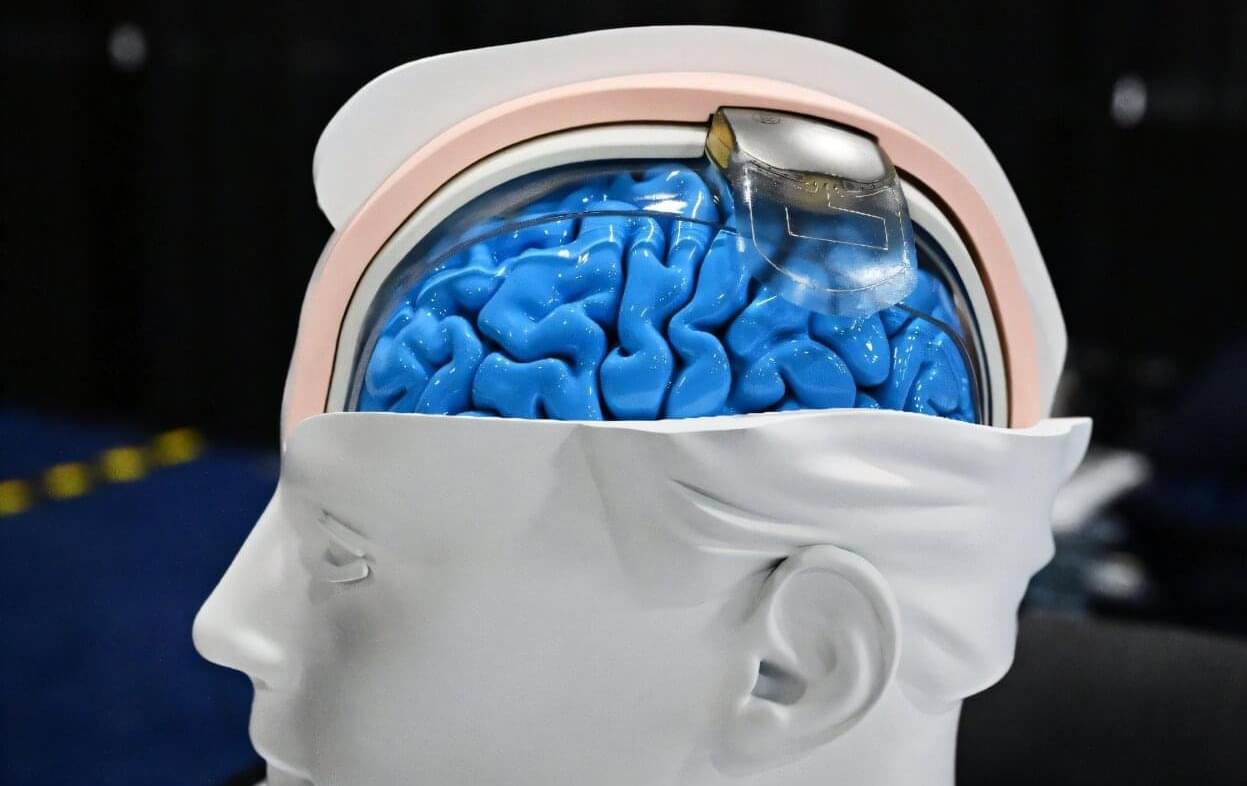
From translating thoughts into words to allowing paralyzed people to walk, the field of neurotechnology has been quietly surging ahead, raising hopes of medical breakthroughs—and profound ethical concerns.
Some observers even think that neurotech could end up being as revolutionary as the far more hyped rise of artificial intelligence (AI).
“People do not realize how much we’re already living in science fiction,” King’s College London researcher Anne Vanhoestenberghe told AFP.
The menopausal transition, or perimenopause, is a 2-to-10-year stretch of hormone irregularity leading up to menopause, when menstruation ceases permanently. It is a time that can come with a variety of challenging symptoms—including hot flashes, sleep disturbances and problems concentrating—as well as often underrecognized mental health challenges, such as heightened irritability, mood swings, increased anxiety and panic attacks. Importantly, a woman’s risk of depression grows two to five times higher than before or after the transition. Suicidal ideation and suicide rates in women are highest between the ages of 45 and 55.
What exactly is happening in the perimenopausal brain to trigger these increased risks? We know that perimenopause represents a major “neurological transition state”; the brain is exposed to drastically changing levels of ovarian hormones, similar to puberty but in reverse. As ovarian function declines, so do levels of the ovarian hormones estradiol and progesterone. The pituitary gland tries to compensate, resulting in erratic hormone fluctuations. These changes are sensed by the ovarian hormone receptors present throughout the brain, particularly in limbic areas important for emotion regulation and memory, such as the hypothalamus and the hippocampus. Neuroimaging studies in living humans have reported changes in estrogen receptor availability and brain metabolism across the menopausal transition.
We also know that estradiol acts as a potent neuromodulator in the brain, affecting multiple neurotransmitter systems, including the serotonergic, noradrenergic and dopaminergic systems, and neuropeptides, such as brain-derived neurotrophic factor—all of which are known to be linked to depression and anxiety disorders. Mouse studies have revealed that hormone shifts across the ovarian cycle induce changes in chromatin organization that underlie changes in hippocampal gene expression, synaptic plasticity and anxiety-related behavior.
It seems likely that the mental health changes observed during perimenopause are related to the shifts in hormones and their receptors in the brain. Few studies, however, have explored the biological mechanisms underlying the increased psychiatric risk. Understanding these changes is essential both for developing new treatments for women in perimenopause who are experiencing mood and memory issues, and for understanding how these changes affect long-term brain health; metabolic changes during perimenopause are thought to increase the risk for Alzheimer’s disease, for instance.
We know little about the mechanisms underlying well-known perimenopause symptoms in the brain. More research—in animals and humans—is essential.
Interested in learning more about tackling the major health challenges of today? Attend our 2025 International Health Lecture: https://bit.ly/3JHOOrp.
Professor Rajesh N Kalaria FMedSci speaks about the global severity of stroke and dementia in society and goes over the findings and lessons from the CogFAST study – cognitive function after stroke study – outcomes of which might mean something for each one of us.
“We can’t change our genes, and we can’t change our age but we can modify every other risk factor for stroke,” he says. He discusses findings that show the impact of modifying some risks factors such as hypertension, and diabetes in elderly post-stroke survivors.
This talk was part of the event \.