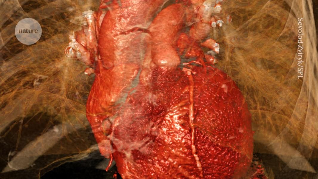
The brain and vagus nerve play a key part in exacerbating tissue damage after a heart attack, but there are ways to block it.
Signs of Sir Terry Pratchett’s dementia may have been present in his writing a decade before his official diagnosis, new research has found. Researchers have examined the lexical diversity—a measure of how varied an author’s word choices are—of 33 books from Pratchett’s Discworld series, focusing specifically on his use of nouns and adjectives.
The study found that Pratchett’s language in “The Lost Continent,” written almost 10 years before his diagnosis of posterior cortical atrophy (PCA), a rare form of Alzheimer’s, showed a significant decline in the complexity of the language used compared to his previous works.
The research team hopes that the study may aid in the early detection of dementia, for which there is currently no cure. The work is published in the journal Brain Sciences.
The study, published in Genomic Psychiatry, identified how stress hormones activate specific RNA molecules called long noncoding RNAs, or IncRNAs, that interact with the gene-silencing complex PRC2, turning off genes that are vital to communication between neurons. In essence, these IncRNAs act like “switches,” turning off functionality for more than 3,000 genes, many of which support neurotransmitter signaling and other processes that are essential for healthy brain functioning. The study specifically discovered 79 IncRNAs that were significantly altered under stress conditions.
While scientists have long understood that stress hormones send signals to the brain that affect gene functionality, it was previously unknown as to exactly how these signals create long-lasting changes inside cells. The study, led by Yogesh Dwivedi, Ph.D., Distinguished Professor and Elesabeth Ridgely Shook Endowed Chair in the Department of Psychiatry and Behavioral Neurobiology, and co-director of UAB Depression and Suicide Center, uncovers how lncRNAs associate with a molecule called polycomb repressive complex 2, or PRC2, to modify chromatin following activation of the glucocorticoid receptor, or GR — the cell’s master regulator of stress response. Chromatin is important in relaying messages from the external environment, including stressful conditions, to alter the genetic composition, a process known as epigenetics.
“As chronic stress is a major risk factor for conditions like major depressive disorder, this newly uncovered link between stress hormones and IncRNA gene silencing could potentially lead to more targeted mental health treatments,” Dwivedi said. “In fact, stress-induced changes in chromatin structure have been implicated in a range of psychiatric and neurodegenerative conditions.”
IncRNAs act like “switches,” turning off functionality for more than 3,000 genes that are essential for healthy brain functioning. A recent groundbreaking study from researchers at the University of Alabama at Birmingham highlights the discovery of a molecular link between stress hormones and changes in brain cell communication, which could open the door for new treatments to address depression and other psychiatric conditions.

Read “” by Myk Eff on Medium.
When a patient in a clinical trial experiences genuine pain relief from an inert sugar pill, something remarkable occurs that contemporary medicine awkwardly labels the placebo effect — a term that simultaneously acknowledges the phenomenon while dismissing it as mere illusion. Yet what if this dismissal represents not scientific rigor but ontological timidity? What if the placebo effect, rather than being a confounding variable to be controlled away, is actually nature’s clearest demonstration of a quantum interface between consciousness and physiology, hiding in plain sight within the very architecture of our clinical trials? The question is not whether belief heals, but what belief actually is when we take seriously the contemporary understanding that information itself possesses physical reality.
The empirical robustness of placebo effects has become impossible to ignore. In their comprehensive meta-analysis published in The Lancet, Hróbjartsson and Gøtzsche (2001) examined 114 clinical trials and found that while placebo effects vary considerably across conditions, they demonstrate genuine clinical significance in pain reduction, with effect sizes rivaling those of established pharmaceutical interventions. More provocatively, Benedetti’s research on placebo analgesia has revealed that the effect operates through identifiable neurochemical pathways — placebo-induced pain relief can be blocked by naloxone, an opioid antagonist, demonstrating that the patient’s belief literally triggers the release of endogenous opioids (Benedetti, Mayberg, Wager, Stohler, & Zubieta, 2005). This is not imagination overriding reality; this is imagination as a physical force, translating expectation into molecular cascade.
Yet the standard neurobiological explanation, while accurate, remains curiously incomplete. Yes, belief activates specific neural circuits; yes, these circuits trigger biochemical responses; yes, measurable physiological changes occur. But this mechanistic account merely pushes the mystery one level deeper. How does the abstract informational content of a belief — the semantic meaning this pill will relieve my pain — couple to the physical substrate of neurons and neurotransmitters? The conventional answer invokes learning, conditioning, and expectation, but these terms describe the phenomenon without explaining the fundamental ontological transition from meaning to matter, from information to effect.

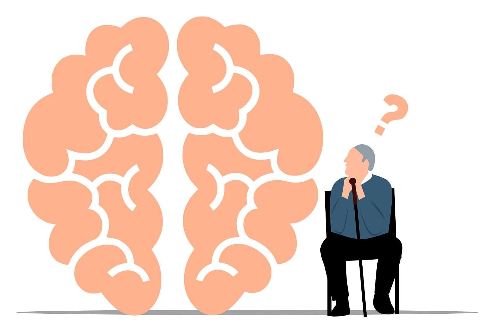
There’s still so much we don’t know about Alzheimer’s disease, but the link between poor sleep and worsening disease is one that researchers are exploring with gusto.
A study published in 2023 found that using sleeping pills to get some shut-eye could reduce the buildup of toxic clumps of proteins in fluid that washes the brain clean every night.
People who took suvorexant, a common treatment for insomnia, for two nights at a sleep clinic experienced a slight drop in amyloid-beta and tau, two proteins that pile up in Alzheimer’s disease.

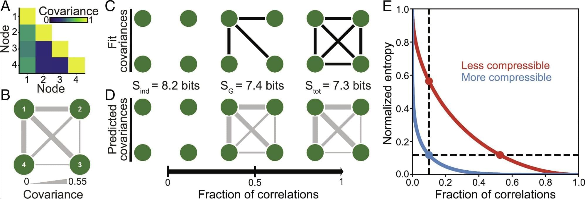
One of the most-viewed PNAS articles in the last week is “Quantifying the compressibility of the human brain.” Explore the article here: https://ow.ly/jGEu50Y6heQ
For more trending articles, visit https://ow.ly/FjuI50Y6heP.
In the human brain, the allowed patterns of activity are constrained by the correlations between brain regions. Yet it remains unclear which correlations—and how many—are needed to predict large-scale neural activity. Here, we present an information-theoretic framework to identify the most important correlations, which provide the most accurate predictions of neural states. Applying our framework to cortical activity in humans, we find that the vast majority of variance in activity is explained by a small number of correlations. This means that the brain is highly compressible: Only a sparse network of correlations is needed to predict large-scale activity. We find that this compressibility is strikingly consistent across different individuals and cognitive tasks and that, counterintuitively, the most important correlations are not necessarily the strongest.