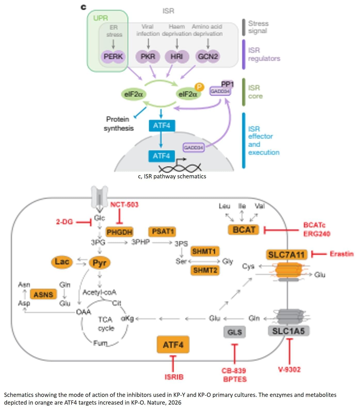
Toggle light / dark theme

The Emergence of AI-Based Wearable Sensors for Digital Health Technology: A Review

The healthcare industry faces a significant shift towards digital health technology, with a growing demand for real-time and continuous health monitoring and disease diagnostics [1, 2, 3]. The rising prevalence of chronic diseases, such as diabetes, heart disease, and cancer, coupled with an aging population, has increased the need for remote and continuous health monitoring [4, 5, 6, 7]. This has led to the emergence of artificial intelligence (AI)-based wearable sensors that can collect, analyze, and transmit real-time health data to healthcare providers so that they can make efficient decisions based on patient data. Therefore, wearable sensors have become increasingly popular due to their ability to provide a non-invasive and convenient means of monitoring patient health. These wearable sensors can track various health parameters, such as heart rate, blood pressure, oxygen saturation, skin temperature, physical activity levels, sleep patterns, and biochemical markers, such as glucose, cortisol, lactates, electrolytes, and pH and environmental parameters [1, 8, 9, 10]. Wearable health technology includes first-generation wearable technologies, such as fitness trackers, smartwatches, and current wearable sensors, and is a powerful tool in addressing healthcare challenges [2].

The data collected by wearable sensors can be analyzed using machine learning (ML) and AI algorithms to provide insights into an individual’s health status, enabling early detection of health issues and the provision of personalized healthcare [6,11]. One of the most significant advantages of AI-based wearable health technology is to promote preventive healthcare. This enables individuals and healthcare providers to proactively address symptomatic conditions before they become more severe [12,13,14,15]. Wearable devices can also encourage healthy behavior by providing incentives, reminders, and feedback to individuals, such as staying active, hydrating, eating healthily, and maintaining a healthy lifestyle by measuring hydration biomarkers and nutrients.

Stem Cell Injection May Soon Reverse Vision Loss Caused By Age-Related Macular Degeneration

Contact: Cara Martinez | Email: cara.martinez@cshs.org

Los Angeles — April 14, 2015 – An injection of stem cells into the eye may soon slow or reverse the effects of early-stage age-related macular degeneration, according to new research from scientists at Cedars-Sinai. Currently, there is no treatment that slows the progression of the disease, which is the leading cause of vision loss in people over 65.

“This is the first study to show preservation of vision after a single injection of induced neural progenitor stem cells into a AMD-like rat model for retinal degeneration,” said Shaomei Wang, MD, PhD, lead author of the study published in the journal STEM CELLS and a research scientist in the Eye Program at the Cedars-Sinai Board of Governors Regenerative Medicine Institute.

Social disadvantage accelerates aging

As an initial step, we selected ARDs associated with hallmarks of aging. These included a total of 83 diseases linked to one or more hallmarks of aging, based on the taxonomy put forward in ref. 4 (Supplementary Table 2). Support for this taxonomy comes from multiple sources. Analyses of electronic health records from general practice and hospitalizations identified more than 200 diseases with incidence rates increasing with chronological age6,22. Researchers linked a subset of these ARDs to specific hallmarks of aging using several approaches: mining 1.85 million PubMed abstracts on human aging, identifying shared genes in the genome-wide association study catalog, conducting gene set enrichment analysis and analyzing disease co-occurrence networks within each hallmark4.

We confirmed the co-occurrence of ARDs within each hallmark in 492,257 participants from the UK Biobank study23. The presence of one ARD increased the risk of developing another ARD related to the same hallmark, with clustering coefficients ranging from 0.76 for LOP-specific ARDs to 0.92 for SCE-specific ARDs. These findings corroborated the hallmark-specific clustering of ARDs (Extended Data Figs. 3 and 4)23.

In time-to-event analyses of UK Biobank and FPS participants without these ARDs at baseline (n ranging from 477,325 to 492,294 in the UK Biobank and from 278,272 to 286,471 in the FPS, depending on the social disadvantage indicator and ARD), social disadvantage—indicated by education and adult SES (neighborhood deprivation)—was associated with a higher risk of developing ARDs. In the UK Biobank, the age-, sex-and ethnicity-adjusted hazard ratio for developing any ARD was 1.31 (95% confidence interval (CI) 1.29–1.33) for individuals with low compared with high education. For individuals with high versus low adult SES, the hazard ratio was 1.21 (95% CI 1.20–1.23). In the FPS, the corresponding hazard ratios were 1.28 (95% CI 1.25–1.31) and 1.23 (95% CI 1.20–1.27), respectively.

Epidermal Growth Factor Stability and Cell Proliferation Enhanced by Antioxidants

1. Introduction.

The natural production of EGF, a short polypeptide hormone, promotes the processes of proliferation, expansion, and division of cells [1]. For in vitro cell culture, EGF functions as a growth factor [2] and has an effective mitogenic effect on endothelial cells, fibroblasts, and most epithelial tissues. Its biological functions rely on associating itself with a specific cell membrane receptor [3]. Because EGF plays a crucial role as a mitogen in the proliferation of various cell types both in vivo and in vitro, it has been used in the therapeutic and cosmetic areas [4] to cover scars and reduce the appearance of aging skin [1]. Moreover, recombinant EGF is used topically for diabetic foot ulcers [5]. The structures and properties of proteins vary; favorable conditions are necessary for conformation, stability, and proper function. In contrast, a protein degrades, denatures, or precipitates when it is exposed to unfavorable conditions or when its natural environment changes suddenly. Recombinant human EGF is most frequently degraded by oxidation and deamidation [6]. These reactions typically have long-term implications. For protein solutions to remain stable and have a longer shelf life, excipients may need to be added, depending on how the protein is used in the experiment and other factors. When it comes to the chemical and physical degradation of proteins, the solution environment plays a crucial role in protein formulations. Of particular concern are buffer types, pH, and antioxidants [7]. Even though antioxidants assist in stability and solubility in liquid solutions, which help to preserve protein structure and function, they are frequently considered inactive ingredients in pharmaceutical compositions [8] [9].

Since an unstable protein solution can impact the product’s appearance, potency, purity, healing effects, and cell proliferation, in vitro protein stabilization is an essential practical consideration for the development of an effective EGF formulation. The stability of EGF in solution has been well documented in several in vivo solutions [10]. Though there have been numerous reports on EGF stability, none have specifically addressed treatment in cell culture conditions. Since it has a big influence on several aspects of the parenteral formulation creation process and EGF-based cell proliferation, the study of EGF stability in cell culture medium has gotten little attention. But since many of these in vitro tests are conducted in non-physiological settings, such as organic solvents or acidic solutions [11], they frequently fail to yield qualitatively positive results in cellular therapies.

Exercise in a Pill? Brown Fat Discovery May Extend Lifespan and Boost Fitness

Rutgers researchers found that increased brown fat improves longevity and exercise capacity in mice. They aim to develop a drug that replicates these benefits in humans.

Rutgers Health researchers have made discoveries about brown fat that could pave the way for helping people stay physically fit as they age.

A team from Rutgers New Jersey Medical School found that mice lacking a specific gene developed an unusually potent form of brown fat tissue, which extended lifespan and increased exercise capacity by approximately 30%. The team is now working on a drug that could replicate these effects in humans.

Advancing light-to-electricity energy conversion: New method extends lifespan of plasmonic hot holes

When light interacts with metallic nanostructures, it instantaneously generates plasmonic hot carriers, which serve as key intermediates for converting optical energy into high-value energy sources such as electricity and chemical energy. Among these, hot holes play a crucial role in enhancing photoelectrochemical reactions. However, they thermally dissipate within picoseconds (trillionths of a second), making practical applications challenging.

Now, a Korean research team has successfully developed a method for sustaining hot holes longer and amplifying their flow, accelerating the commercialization of next-generation, high-efficiency, light-to-energy conversion technologies.

The research team, led by Distinguished Professor Jeong Young Park from the Department of Chemistry at KAIST, in collaboration with Professor Moonsang Lee from the Department of Materials Science and Engineering at Inha University, has successfully amplified the flow of hot holes and mapped local current distribution in real time, thereby elucidating the mechanism of photocurrent enhancement. The work is published in Science Advances.

Who does Spiderman vote for? Study shows people project their political views onto fictional heroes and villains

From a very young age, we’re socialized to view the world as being made up of “goodies” and “baddies.” When you’re a child fooling around with your friends in the playground, nobody ever wants to be the baddy. And when it comes to dressing up, everybody wants to be Luke Skywalker—not Darth Vader.

This oversimplified way of viewing the world as being made up of right and wrong or good people and bad people doesn’t dissipate as we grow older. If anything, it tends to solidify as we form the that define who we are in adult life.

This is particularly the case when it comes to our political identities and, specifically, the partisan identities and loyalties that individuals attach themselves to.

Endothelial TDP-43 depletion disrupts core blood–brain barrier pathways in neurodegeneration

The study, “Endothelial TDP-43 Depletion Disrupts Core Blood-Brain Barrier Pathways in Neurodegeneration,” was published on March 14, 2025. The lead author, Omar Moustafa Fathy, an MD/Ph. D. candidate at the Center for Vascular Biology at UConn School of Medicine, conducted the research in the laboratory of senior author Dr. Patrick A. Murphy, associate professor and newly appointed interim director of the Center for Vascular Biology. The study was carried out in collaboration with Dr. Riqiang Yan, a leading expert in Alzheimer’s disease and neurodegeneration research.

This work provides a novel and significant exploration of how vascular dysfunction contributes to neurodegenerative diseases, exemplifying the powerful collaboration between the Center for Vascular Biology and the Department of Neuroscience. While clinical evidence has long suggested that blood-brain barrier (BBB) dysfunction plays a role in neurodegeneration, the specific contribution of endothelial cells remained unclear. The BBB serves as a critical protective barrier, shielding the brain from circulating factors that could cause inflammation and dysfunction. Though multiple cell types contribute to its function, endothelial cells—the inner lining of blood vessels—are its principal component.

“It is often said in the field that ‘we are only as old as our arteries’. Across diseases we are learning the importance of the endothelium. I had no doubt the same would be true in neurodegeneration, but seeing what these cells were doing was a critical first step,” says Murphy.

Omar, Murphy, and their team tackled a key challenge: endothelial cells are rare and difficult to isolate from tissues, making it even harder to analyze the molecular pathways involved in neurodegeneration.

To overcome this, they developed an innovative approach to enrich these cells from frozen tissues stored in a large NIH-sponsored biobank. They then applied inCITE-seq, a cutting-edge method that enables direct measurement of protein-level signaling responses in single cells—marking its first-ever use in human tissues.

This breakthrough led to a striking discovery: endothelial cells from three different neurodegenerative diseases—Alzheimer’s disease (AD), amyotrophic lateral sclerosis (ALS), and frontotemporal dementia (FTD)—shared fundamental similarities that set them apart from the endothelium in healthy aging. A key finding was the depletion of TDP-43, an RNA-binding protein genetically linked to ALS-FTD and commonly disrupted in AD. Until now, research has focused primarily on neurons, but this study highlights a previously unrecognized dysfunction in endothelial cells.

“It’s easy to think of blood vessels as passive pipelines, but our findings challenge that view,” says Omar. “Across multiple neurodegenerative diseases, we see strikingly similar vascular changes, suggesting that the vasculature isn’t just collateral damage—it’s actively shaping disease progression. Recognizing these commonalities opens the door to new therapeutic possibilities that target the vasculature itself.”

How long will you live? New evidence says it’s much more about your choices than your genes

One of the most enduring questions humans have is how long we’re going to live. With this comes the question of how much of our lifespan is shaped by our environment and choices, and how much is predetermined by our genes.

A study recently published in the prestigious journal Nature Medicine has attempted for the first time to quantify the relative contributions of our environment and lifestyle versus our genetics in how we age and how long we live.

The findings were striking, suggesting our environment and lifestyle play a much greater role than our genes in determining our longevity.