Thanks to CRISPR, medical specialists will soon have unprecedented control over how they treat and prevent some of the most challenging genetic disorders and diseases.
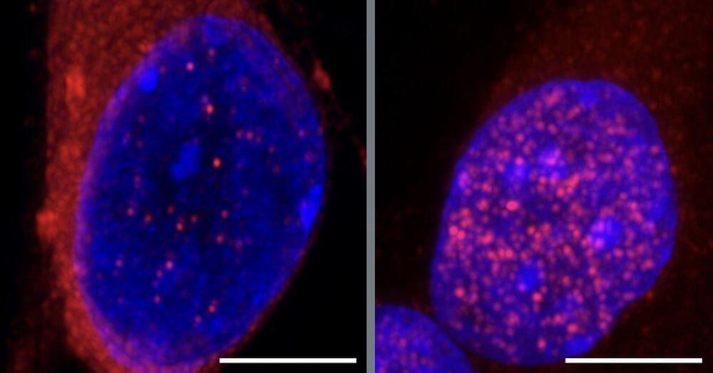
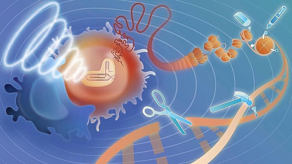
CRISPR (Clustered Regularly Interspaced Short Palindromic Repeats) is a Nobel Prize-winning gene-editing tool, already widely used by scientists to cut and modify DNA sequences to turn genes on and off or insert new DNA that can correct abnormalities. CRISPR uses an enzyme known as Cas9 to cut and alter DNA.
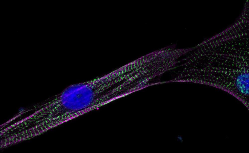
Engineers at the USC Alfred E. Mann Department of Biomedical Engineering have now developed an update to the tool that will allow CRISPR technology to be even more powerful with the help of focused ultrasound.