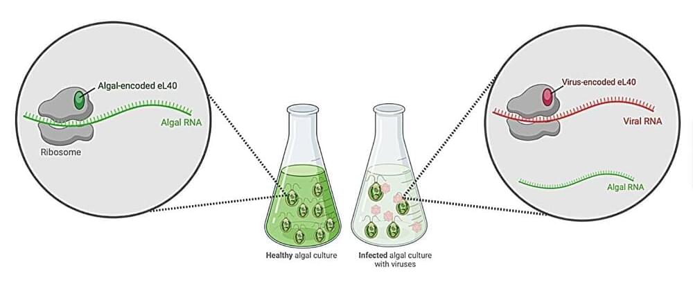
Toggle light / dark theme

Researchers discover possible new treatment for triple-negative breast cancer

Zachary Schug, Ph.D., assistant professor in the Molecular and Cellular Oncogenesis Program of the Ellen and Ronald Caplan Cancer Center at The Wistar Institute, has published a new paper in the journal Nature Cancer. Schug’s paper, titled “Acetate acts as a metabolic immunomodulator by bolstering T-cell effector function and potentiating antitumor immunity in breast cancer,” demonstrates a double-acting mechanism for fighting a particularly aggressive, difficult-to-treat form of breast cancer. Schug’s research shows how silencing a certain gene, ACSS2, may improve existing treatments for patients.

Triple-negative , or TNBC, affects 10–15% of patients with breast cancer in the US. TNBC is called “triple-negative” because the cancer lacks an , a , and a HER2 (human epidermal growth factor) receptor. The absence of any of these receptors—receptors that when present in other forms of breast cancer, can be effectively targeted during treatment—makes treating TNBC quite difficult, and patients with TNBC have limited treatment options.

TNBC’s notorious aggression makes the technical challenge of finding a reliably effective treatment target all the more serious: compared to other breast cancers, TNBC grows faster and resists treatment more stubbornly. All these factors contribute to the fact that TNBC patients suffer from worse prognoses.

A modern digital light processing technology to 3D print microfluidic chips

Conventional manufacturing methods such as soft lithography and hot embossing processes can be used to bioengineer microfluidic chips, albeit with limitations, including difficulty in preparing multilayered structures, cost-and labor-consuming fabrication processes as well as low productivity.

Materials scientists have introduced digital light processing as a cost-effective microfabrication approach to 3D print microfluidic chips, although the fabrication resolution of these microchannels are limited to a scale of sub-100 microns.

In a new report published in Microsystems and Nanoengineering, Zhuming Luo and a scientific team in , and chemical engineering in China developed an innovative digital light processing method.

Innovative Gene Screening in Human Tissue May Unlock Autism’s Secrets

Summary: Researchers pioneered a groundbreaking method called “CHOOSE” to investigate genes tied to autism spectrum disorder (ASD) within human tissue. This technique allows for simultaneous examination of key transcriptional regulator genes linked to autism in a single organoid.

Utilizing CHOOSE, the team pinpointed mutations in 36 genes known to heighten autism risk, shedding light on how they influence brain development. The revelations from these organoids mirrored clinical observations, underscoring the potential of this method in advancing our understanding of neurodevelopmental disorders.

AI now used in the fight against global infectious diseases

Our hope is for COVID-19 to never repeat itself,’ said the new program’s executive director.

A program run by a Canadian university is seeking to improve global health care for the most vulnerable by examining how artificial intelligence (AI) can enhance readiness for infectious disease epidemics in the Global South.

This is according to a report by CTV News published on Wednesday.


Interesting Engineering is a cutting edge, leading community designed for all lovers of engineering, technology and science.

The surprising origin of a deadly hospital infection

Hospital staff spend a significant amount of time working to protect patients from acquiring infections while they are being cared for in the hospital. They employ various methods from hand hygiene to isolation rooms to rigorous environmental sanitation. Despite these efforts, hospital-onset infections still occur—the most common of which is caused by the bacterium Clostridioides difficile, or C. diff, the culprit of almost half a million infections in the U.S. each year.

Surprising findings from a study in Nature Medicine suggest that the burden of C. diff infection may be less a matter of hospital transmission and more a result of characteristics associated with the themselves.

The study team, led by Evan Snitkin, Ph.D. and Vincent Young, M.D., Ph.D., both members of the Departments of Microbiology & Immunology and Internal Medicine/Infectious Diseases at University of Michigan Medical School and Mary Hayden, M.D. of Rush University Medical Center, leveraged ongoing epidemiological studies focused on hospital-acquired infections that enabled them to analyze daily fecal samples from every patient within the at Rush University Medical Center over a nine-month period.

Forward genetic screening using fundus spot scale identifies an essential role for Lipe in murine retinal homeostasis

Year 2023 😗


Data from patients with AMD, retinal dystrophies, and diabetic retinopathy indicate an important role of immune cells, including microglia, in the pathogenesis of these retinal diseases1. The accumulation of drusen components provides an environment rich in chemoattractants for microglia and leads to their translocation to the subretinal space in AMD2,4. The involvement of microglia in the activation of the NLRP3 inflammasome and the promotion of proinflammatory cytokine secretion has been confirmed in in vitro and animal studies11,12,14. In patients with retinal dystrophies like retinitis pigmentosa, it has been shown that microglia become activated in response to signals from degenerating rod photoreceptors and migrate to the outer retinal layers4. There, they participate in the phagocytosis of debris and dying cells and secrete proinflammatory factors. Mouse models of retinal degeneration (e.g. rd1, rd7, rd8, and rd10 models) confirm many of these conclusions9,10,13,15, but make it clear that the role of microglia may also be homeostatic, depending on both stimuli and anatomical location within the retina7,20. Activated microglia are observed at all the stages of human diabetic retinopathy3,8 and also feature prominently in many animal models of the disease44,45. Finally, accumulations of activated microglia are also seen in a variety of animal models of retinal degeneration, including light-induced retinal degeneration and models based on complement dysregulation34,46,47.

The pathways regulating immune surveillance, cell trafficking, and neuroinflammation in the retina are not well understood. A large number of molecules and processes have been implicated, ranging from chemokines involved in chemotaxis, cytokines involved in activation, factors that regulate oxidative stress and complement activation, and immunoregulatory proteins. In such a complex biological system, the unbiased nature of a forward genetics approach is particularly valuable in identifying genes affecting these immune cell processes. Furthermore, the accumulation of subretinal microglia, visible as or correlated with the accumulation of fundus spots, can serve as a marker for retinal pathology and thus as a screen for genes essential to retinal homeostasis. Our approach here has two important advantages relative to all prior forward genetics studies of the retina: 1. We are systematically applying a semiquantitative fundus spot scale to fundus photographs, and 2. Our pipeline is the only one in which all mice screened are G3 mice that have been pre-genotyped at all mutant loci. Our unbiased identification of 6 gene-phenotype associations to retinal pathology with strong literature support using our fundus spot scale screen is proof of concept supporting the efficacy of our approach. We identified other associations that had not been reported in the literature at the time of the screening. From those, we first concentrated our efforts on the gene Lipe, partly because the fundus spot scale was the only parameter leading to its identification.

In order to confirm our findings in ENU-mutagenized mice and also to explore the role of Lipe in retinal homeostasis, a CRISPR-generated Lipe−/− mouse line was generated. Imaging of the retinas on these mice confirmed an early and prominent accumulation of fundus spots. Furthermore, we found a similar widespread accumulation of hyperautofluorescent spots in these mice. We were also able to show that Lipe−/− mice have increased accumulation of subretinal Iba1+/CD16+/TMEM119+/CCR2− cells consistent with activated microglia. It can be argued that microglia migrating to the subretinal space are by definition showing some level of activation48,49,50. But our findings of well-accepted morphological signs of activation and co-staining with CD16, a marker of microglial activation10,34,51,52, further support this conclusion.

Potential Therapeutic Benefit of NAD+ Supplementation for Glaucoma and Age-Related Macular Degeneration

Year 2020 Immortality in eyes achieved with Nad plus 😗😁😘.


Glaucoma and age-related macular degeneration are leading causes of irreversible blindness worldwide with significant health and societal burdens. To date, no clinical cures are available and treatments target only the manageable symptoms and risk factors (but do not remediate the underlying pathology of the disease). Both diseases are neurodegenerative in their pathology of the retina and as such many of the events that trigger cell dysfunction, degeneration, and eventual loss are due to mitochondrial dysfunction, inflammation, and oxidative stress. Here, we critically review how a decreased bioavailability of nicotinamide adenine dinucleotide (NAD; a crucial metabolite in healthy and disease states) may underpin many of these aberrant mechanisms. We propose how exogenous sources of NAD may become a therapeutic standard for the treatment of these conditions.

Researchers create optical device that can kill pathogens on surfaces while remaining safe for humans

While it has long been known that ultraviolet (UV) light can help kill disease-causing pathogens, the COVID-19 pandemic has put a spotlight on how these technologies can rid environments of germs. However, the excimer lamps and LEDs that can directly emit light in the required deep-UV wavelengths generally have low efficiency or suffer from short lifetimes. Moreover, UV light of the wrong wavelength can actually be harmful to human cells.

Now, a team led by researchers from Osaka University has shown how an made of can be used to generate deep-UV light in a method wholly different from previous approaches. The team made use of a process called “second harmonic generation,” which relies on the fact that the frequency of a photon, or particle of light, is proportional to its energy. The study is published in the journal Applied Physics Express.

Most are considered “linear” with respect to their response to light, i.e., photons cannot interact with each other. However, inside certain “nonlinear” materials, two photons can be combined into a with twice the energy, and thus, twice the frequency.