Toggle light / dark theme

But are you ready to explain this complex disease in terms that your child can understand?

Keep It Simple for Young Children Use language that is appropriate for your child’s age to describe what happens when asthma flares-up. Try to avoid medical terminology and details that will confuse a young child. Stick to the basic information at first. This includes making your child aware of what triggers symptoms and giving very basic understanding of what happens to cause the symptoms. Visit Just for Kids for story books, games and puzzles that explain asthma in terms your child can understand.

Medication management is not necessarily an issue with young children, as it is your responsibility as a caregiver to make certain you give your child medication as prescribed. Set a routine so it’s easy to remember when these medications should be taken. Practice with a nebulizer, peak flow meter and spacer so that your child understands how each device helps manage asthma.

150 YEARS MAXIMUM BIOLOGICAL AGE — “We observed, that the age-dependent population DOSI distribution broadening could be explained by a progressive loss of physiological resilience measured by the DOSI auto-correlation time. Extrapolation of this trend suggested that DOSI recovery time and variance would simultaneously diverge at a critical point of 120 − 150 years of age corresponding to a complete loss of resilience. The observation was immediately confirmed by the independent analysis of correlation properties of intraday physical activity levels fluctuations collected by wearable devices. We conclude that the criticality resulting in the end of life is an intrinsic biological property of an organism that is independent of stress factors and signifies a fundamental or absolute limit of human lifespan.”


We investigated the dynamic properties of the organism state fluctuations along individual aging trajectories in a large longitudinal database of CBC measurements from a consumer diagnostics laboratory. To simplify the analysis, we used a log-linear mortality estimate from the CBC variables as a single quantitative measure of aging process, henceforth referred to as dynamic organism state index (DOSI). We observed, that the age-dependent population DOSI distribution broadening could be explained by a progressive loss of physiological resilience measured by the DOSI auto-correlation time. Extrapolation of this trend suggested that DOSI recovery time and variance would simultaneously diverge at a critical point of 120 − 150 years of age corresponding to a complete loss of resilience. The observation was immediately confirmed by the independent analysis of correlation properties of intraday physical activity levels fluctuations collected by wearable devices. We conclude that the criticality resulting in the end of life is an intrinsic biological property of an organism that is independent of stress factors and signifies a fundamental or absolute limit of human lifespan.

P.O. Fedichev is a shareholder of Gero LLC. A.Gudkov is a member of Gero LLC Advisory Board. T.V. Pyrkov, K. Avchaciov, A.E. Tarkhov, L. Menshikov, and P.O. Fedichev are employees of Gero LLC.

In the biomedical field, optical characterization of cells and tissues is a valuable tool for understanding physiological mechanisms. Current biomedical optical imaging techniques include fluorescence imaging [1], confocal microscopy [2], optical coherence tomography [3], two-photon microscopy [4], near-infrared spectroscopy [5], and diffuse optical tomography [6]. These techniques have significantly advanced biomedical technology and are widely used for both preclinical and clinical purposes. However, the strong optical scattering within turbid biological tissues fundamentally limits the imaging depth of these pure optical imaging techniques to no deeper than the optical ballistic depth ( 1 mm). Thus, their observation depth is superficial and other imaging modalities are needed to explore deeper layers of biological tissue [7].

Photoacoustic imaging (PAI), a promising biomedical technique, achieves superior imaging depths by forming images from optically-derived acoustic signals, which inherently attenuate less than optical signals in biological tissue [8, 9, 10]. PAI is based on the photoacoustic (PA) effect, in which energy is converted from light to acoustic waves via thermoelastic expansion [11,12,13,14,15,16]. To generate PA waves, a laser beam with a typical pulse width of a few nanoseconds illuminates the target tissue. The optical chromophores in biological tissue absorb the light energy and then release the energy soon after. The energy release can can occur as either light energy with a slightly shifted wavelength or as thermal energy that causes thermoelastic expansion. In PAI, the rapidly alternating thermoelastic expansion and contraction caused by pulsed light illumination generates vibrations in tissue that propagate as acoustic waves called PA waves. The generated PA waves can be detected by conventional ultrasound (US) transducers for image generation. Because PAI and ultrasound imaging (USI) share the same signal reception and image reconstruction principle, the two modalities are technically fully compatible and can be implemented in a single US imaging platform accompanied with pulse laser source [17,18,19,20,21]. Since PAI can capture the photochemical properties of the target site, combining PAI with USI can provide both chemical and structural information about a target tissue.

One distinctive advantage of PAI is that its resolution and imaging depth can be adjusted to suit a specific target area. The resolution of PA signals depends on both the optical focus of the excitation laser and the acoustic focus of the receiving US transducer [22], so images with tuned spatial resolutions and imaging depths can be achieved by modifying the system configuration [23]. PAI’s wide applications to date have included nanoscale surface and organelle imaging [24,25,26,27,28], microscale cellular imaging [29,30,31,32], macroscale small animal imaging [33,34,35], and clinical human imaging [36,37,38].

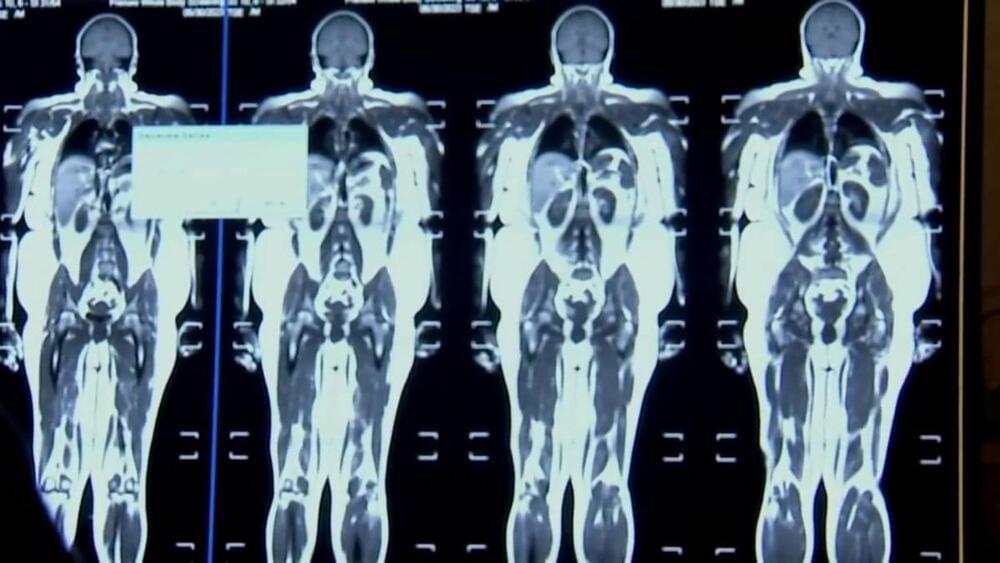
A new study finds artificial intelligence could help predict pancreatic cancer. Dr. Chris Sander, one of the co-authors of the study, joined CBS News to talk about the findings.

#news #ai #cancer.

CBS News Streaming Network is the premier 24/7 anchored streaming news service from CBS News and Stations, available free to everyone with access to the Internet. The CBS News Streaming Network is your destination for breaking news, live events and original reporting locally, nationally and around the globe. Launched in November 2014 as CBSN, the CBS News Streaming Network is available live in 91 countries and on 30 digital platforms and apps, as well as on CBSNews.com and Paramount+.

Watch CBS News: https://cbsn.ws/1PlLpZ7c.

X-ray technology plays a vital role in medicine and scientific research, providing non-invasive medical imaging and insight into materials. Recent advancements in X-ray technology enable brighter, more intense beams and imaging of increasingly intricate systems in real-world conditions, like the insides of operating batteries.

To support these advancements, scientists are working to develop X-ray materials that can withstand bright, high-energy X-rays—especially those from large X-ray synchrotrons—while maintaining sensitivity and cost-effectiveness.

A team of scientists at the U.S. Department of Energy’s (DOE) Argonne National Laboratory and their colleagues have demonstrated exceptional performance of a new material for detecting high energy X-ray scattering patterns. With excellent endurance under ultra-high X-ray flux and relatively low cost, the detector material may find wide application in synchrotron-based X-ray research.

Researchers at ETH Zurich recently identified a previously unknown compartment in mammalian cells. They have named it the exclusome. It is made up of DNA rings known as plasmids. The researchers have published details of their discovery in the journal Molecular Biology of the Cell.

The new compartment is in the cell plasma; it is previously uncharacterized in the literature. It is exceptional because eukaryotic cells (cells with nuclei) usually keep most of their DNA in the , where it is organized into chromosomes.

Some of the plasmids that end up in the exclusome originate from outside the cell, while others—known as telomeric rings—come from the capped ends of chromosomes, the telomeres. Particularly in certain , the ones from the telomeres are regularly pinched off and joined together to form rings. However, these don’t contain the blueprints for proteins.

A team led by Professor Choi Hong-Soo in the Department of Robotics and Mechatronics Engineering at DGIST has discovered a method to enhance the penetration of magnetic nanoparticles into cancer cells and their magnetic hyperthermia effects through research on chain disassembly and magnetic propulsion mechanisms using a rotational magnetic field.

Published in the journal ACS Nano, their study focused on the delivery of magnetic therapeutic agents using magnetic fields, an area receiving attention in the field of cancer treatment. It is expected to contribute significantly by improving drug delivery efficiency and therapeutic effects in targeted cancer treatments.

Recently, the development of targeted therapeutics that selectively treat has been gaining attention in the field of cancer treatment. Among them, research on magnetic carriers that target cancer cells using magnetic fields is underway. However, a problem arises when magnetic nanoparticles are exposed to a uniform magnetic field with a general form; they form long chains in the direction of the magnetic field, making penetration into cancer cells or tumors difficult and reducing the therapeutic efficacy.

George Church at his most optimistic. June 1, 2022.


Dr George Church talks about combination therapies for age reversal, recently published papers from his lab and expresses his wish on developing inexpensive gene therapies like vaccine that can be equitably distributed to human.

Dr George Church is the Robert Winthrop Professor of Genetics at Harvard Medical School, a Professor of Health Sciences and Technology at Harvard and the Massachusetts Institute of Technology (MIT), and a core faculty member of the Wyss Institute.

Join us on Patreon! https://www.patreon.com/MichaelLustgartenPhD

Discount Links:
At-Home Metabolomics: https://www.iollo.com?ref=michael-lustgarten.
Use Code: CONQUERAGING At Checkout.

NAD+ Quantification: https://www.jinfiniti.com/intracellular-nad-test/
Use Code: ConquerAging At Checkout.

Epigenetic Testing: https://trudiagnostic.com/?irclickid=U-s3Ii2r7xyIU-LSYLyQdQ6…M0&irgwc=1

The human brain can change – but usually only slowly and with great effort, such as when learning a new sport or foreign language, or recovering from a stroke. Learning new skills correlates with changes in the brain, as evidenced by neuroscience research with animals and functional brain scans in people. Presumably, if you master Calculus 1, something is now different in your brain. Furthermore, motor neurons in the brain expand and contract depending on how often they are exercised – a neuronal reflection of “use it or lose it.”

People may wish their brains could change faster – not just when learning new skills, but also when overcoming problems like anxiety, depression and addictions.

… More


Change in the brain usually comes with plenty of effort over time. Neuroscientists are working to understand how psychedelic drugs provide a shortcut that seems to rely on existing brain systems.