Toggle light / dark theme

Biomedical engineers at Duke University have developed a new method to improve the effectiveness of machine learning models. By pairing two machine learning models, one to gather data and one to analyze it, researchers can circumvent limitations of the technology without sacrificing accuracy.

This new technique could make it easier for researchers to use machine learning algorithms to identify and characterize molecules for use in potential new therapeutics or other materials.

The research is published in the journal Artificial Intelligence in the Life Sciences.

The type I CRISPR protein Cas3 works like Pac-Man, chomping away at a continuous stream of nucleotides with intrinsic activity for introducing targeted large deletions from a few hundred base pairs to as large as 200 kb. However, without the molecular equivalent to the four colored ghosts who chase and capture Pac-Man, the broad and unidirectional genome editing activity of Cas3 is essentially unregulated.

Yan Zhang, PhD, assistant professor in the department of biological chemistry at the University of Michigan Medical School, and her collaborators at Cornell University identified two anti-CRISPR proteins that can “turn off” Cas3, paving the way toward safer and better-controlled CRISPR applications.

The research article, “Exploiting activation and inactivation mechanisms in type I-C CRISPR-Cas3 for genome-editing applications,” was published in Molecular Cell.

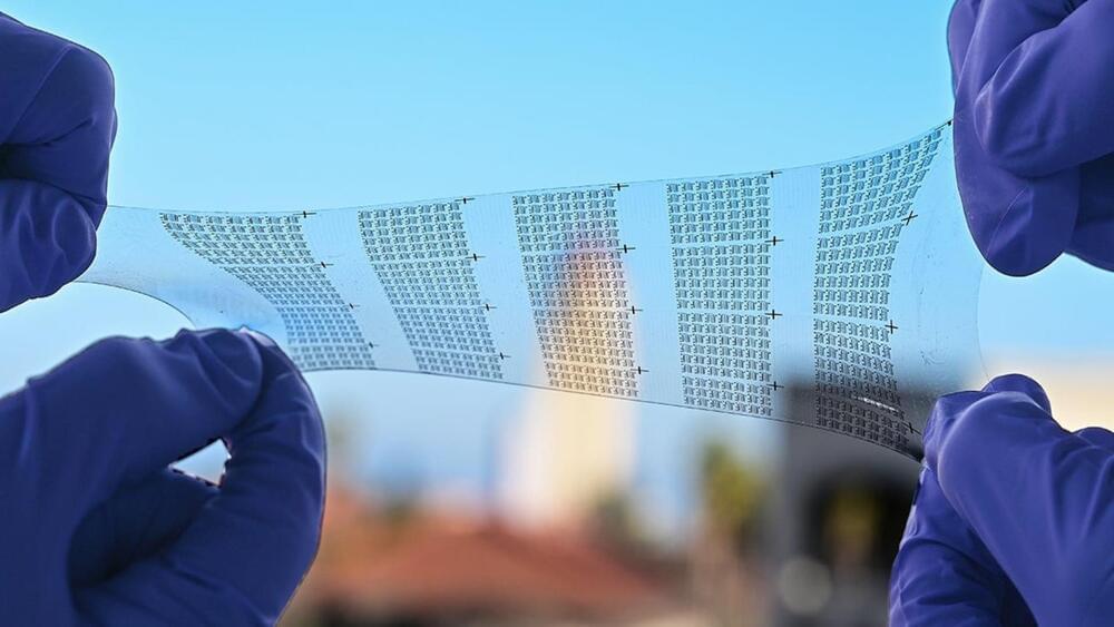
Researchers have created a novel technology utilizing meta-optical devices for thermal imaging. This method offers more detailed information about the objects being imaged, potentially expanding thermal imaging applications in autonomous navigation, security, thermography, medical imaging, and remote sensing.

“Our method overcomes the challenges of traditional spectral thermal imagers, which are often bulky and delicate due to their reliance on large filter wheels or interferometers,” said research team leader Zubin Jacob from Purdue University. “We combined meta-optical devices and cutting-edge computational imaging algorithms to create a system that is both compact and robust while also having a large field of view.”

In Optica, Optica Publishing Group’s journal for high-impact research, the authors describe their new spectro-polarimetric decomposition system, which uses a stack of spinning metasurfaces to break down thermal light into its spectral and polarimetric components. This allows the imaging system to capture the spectral and polarization details of thermal radiation in addition to the intensity information that is acquired with traditional thermal imaging.

Scientific investigations on the ISS’s latest resupply mission include advancements in 3D metal printing, semiconductor manufacturing, reentry thermal protection, robotic surgery, and cartilage tissue regeneration. These studies aim to enhance space mission sustainability and have significant implications for Earth-based technologies and health care.

Tests of a 3D metal printer, semiconductor manufacturing, and thermal protection systems for reentry to Earth’s atmosphere are among the scientific investigations that NASA and international partners are launching to the International Space Station on Northrop Grumman’s 20th commercial resupply services mission. The company’s Cygnus cargo spacecraft is scheduled to launch on a SpaceX Falcon 9 rocket from Cape Canaveral Space Force Station in Florida by late January.

Read more about some of the research making the journey to the orbiting laboratory:

SynapShot, developed by an international research team, marks a major advancement in neuroscience by enabling real-time, live observation of synaptic changes in the brain.

The human brain contains approximately 86 billion neurons and 600 trillion synapses that exchange signals between the neurons to help us control the various functions of the brain including cognition, emotion, and memory. Interestingly, the number of synapses decrease with age or as a result of diseases like Alzheimer’s, and research on synapses thus attracts a lot of attention. However, limitations have existed in observing the dynamics of synapse structures in real-time.

It has been known for more than century that increases in neural activity in the brain drive changes in local blood flow, known as neurovascular coupling. The colloquial explanation for these increases in blood flow (referred to as functional hyperemia) in the brain is that they serve to supply the needs of metabolically active neurons. However, there is an large body of evidence that is inconsistent with this idea. In most cases, baseline blood flow is adequate to supply even elevated neural activity. Neurovascular coupling is irregular, absent, or inverted in many brain regions, behavioral states, and conditions. Increases in respiration can generate increases in brain oxygenation independently of flow changes. Simulations have shown that areas with low blood flow are inescapable and cannot be removed by functional hyperemia given the architecture of the cerebral vasculature. What physiological purpose might neurovascular coupling serve? Here, we discuss potential alternative functions of neurovascular coupling. It may serve supply oxygen for neuromodulator synthesis, to regulate cerebral temperature, signal to neurons, stabilize and optimize the cerebral vascular structure, deal with the non-Newtonian nature of blood, or drive the production and circulation of cerebrospinal fluid around and through the brain via arterial dilations. Understanding the ‘why’ of neurovascular coupling is an important goal that give insight into the pathologies caused by cerebrovascular disfunction.

Like all energy demanding organs, the brain is highly vascularized. When presented with a sensory stimulus or cognitive task, increases in neural activity in many brain regions are accompanied by local dilation of arterioles and other microvessels, increasing local blood flow, volume and oxygenation. The increase in blood flow in response to increased neural activity (known as functional hyperemia) is controlled by a multitude of different signaling pathways via neurovascular coupling (reviewed in [1,2]). These vascular changes can be monitored non-invasively in humans and other species, with techniques (like BOLD fMRI) that are cornerstones in modern neuroscience [3,4]. Chronic disruptions of neurovascular coupling have adverse health effects on the brain. Stress affects neurovascular coupling [5,6], and many neurodegenerative diseases are marked by vascular dysfunction [7].

As one of the few schools on Long Island offering the AP Capstone program in their curricula, Patchogue-Medford High School provides various STEM opportunities for its students.

From Brookhaven National Laboratory to Stony Brook University right at our fingertips, the possibilities are endless. For instance, on December 8 th, four AP Research students joined Dr. Gatz in spending the day at Cold Spring Harbor Laboratory to conduct the necessary experiments for their research papers.

Senior, Carlo Costigliola, and Junior, Isaac Varghese, have decided to focus their research on DNA barcoding. According to International Barcode of Life, “DNA barcoding is a method of specimen identification using short, standardized segments of DNA.”

In 1,868, Jean-Martin Charcot, a neurologist at the Hôpital de Salpétrière in France, first coined the disease “la sclérose en plaques,” which means multiple sclerosis (MS) — to distinguish it from another type of movement disorder later known as Parkinson’s disease.

Though described in 1,868, the cause of MS puzzled scientists for more than a century. This is until a 2022 breakthrough study finally enlightens us that the cause is, oddly, the seemingly innocent Epstein-Barr virus (EBV), a common childhood virus that causes typical fever and sore throat.

Let’s see how one study single-handedly proves what we thought couldn’t be proved; how one study truly deserves to be called a breakthrough; and how thorough and near-perfect science is done.