Toggle light / dark theme

A McGill University-led research collaboration has achieved a breakthrough in understanding how cancer spreads. A clinical study of ovarian and colorectal cancer patients found cancer cells move in the bloodstream in clusters more commonly than was previously thought. The discovery could help doctors more quickly identify which cancer patients are at high risk of having their cancer spread to other organs, knowledge that could guide treatment decisions. The findings also potentially open new avenues for treatment.

The study, published in Communications Medicine, was conducted with researchers and clinicians Anne-Marie Mes-Masson and Dr. Diane Provencher at the Centre hospitalier de l’Université de Montréal, Dr. Peter Metrakos at the Research Institute of the McGill University Health Centre and Luke McCaffrey at the McGill-affiliated Rosalind and Morris Goodman Cancer Institute.

Cancer is responsible for about 1 in 4 deaths in Canada. In most cases, it is not the original tumor that proves fatal, but the cancer spreading to other organs, a process called metastasis. This occurs when circulating tumor cells (CTCs) break away from tumors, enter the bloodstream, and seed new tumors elsewhere in the body. On rare occasions, CTCs break away as a group of cells sticking to one another and forming a cluster.

The strong links between changes in astrocyte structure and function in the context of neurodevelopment and disease have been supported by studies examining astrocyte cytoskeletal markers such as glial fibrillary acidic protein (GFAP) in disease models and postmortem human brain tissue, where increases or decreases in its expression in various brain nuclei are often linked with neurocognitive and psychiatric disorders. Hence, changes in GFAP expression are often the first-line test for astrocyte involvement in disease and support a role for astrocyte dysfunction in major depression, schizophrenia, alcohol and substance use disorders, anorexia nervosa, and bipolar disorder (719), where changes in astrocyte structure, density, complexity, and/or blood vessel association are linked with disrupted astrocyte function. Although reactive astrogliosis remains the single most studied astrocytic response involving morphological adaptations and changes in GFAP expression (20, 21), in recent years, astrocyte morphological plasticity has been shown to be more nuanced. GFAP expression is dynamic across the circadian cycle (2224) and increases with physical exercise and environmental enrichment (25, 26). Moreover, in aging, astrocytes increase or decrease their GFAP expression in different brain regions (27, 28), suggesting heterogeneity in astrocyte form and function.

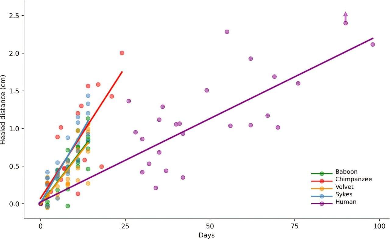
We previously found a notable relationship between astrocyte structure and vulnerability to substance use disorders, with astrocytes in the nucleus accumbens (NAc) altering their association with different neural subcircuits to drive or suppress drug-seeking behavior depending on heroin availability (2931). The NAc is critical for regulating behavioral outputs in response to rewards, including substances of abuse and natural reinforcers, such as food or sucrose. The NAc is composed of core and shell subregions that are themselves heterogeneous structures with regard to synaptic input and output connectivity and function (3236). Heterogeneity has been observed in astrocyte morphology within the NAc core (3, 30, 37), but studies have not yet examined how astrocyte structure and function differ across NAc subregions at baseline or in response to operant conditioning with natural or pathological reinforcers.

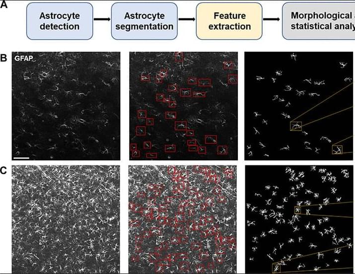
To address this gap, we developed an automated pipeline for single-cell morphological analysis of astrocytes that integrates state-of-the-art deep learning models for astrocyte detection and segmentation, together with highly sensitive geometrical tools for precise quantitation of single-cell morphological characteristics. We introduce the rigorous notion of morphological distance (MD) to measure alterations in astrocyte morphology and compare astrocyte subpopulations according to their structural characteristics. By applying this pipeline in combination with supervised machine learning, we found that single-astrocyte morphological characteristics were predictive not only of anatomical location within the NAc at baseline but also of the availability of heroin or sucrose at the moment of image capture. This geometrically sensitive approach yields substantially more detailed information about astrocyte structure than previously applied manual or semiautomated approaches and serves as a rigorous quantitative assay for identifying brain nuclei where astrocytes undergo plasticity in the context of disease. We found that astrocyte structural plasticity across the NAc was disrupted in animals that had been exposed to heroin but not sucrose, consistent with a largely protective role for NAc astrocytes in maintaining synaptic homeostasis and behavioral flexibility. We also found that astrocyte structural plasticity in the dorsomedial portion of the NAc shell was uniquely engaged during the initiation of opioid but not sucrose seeking, suggesting the involvement of this structure in drug relapse.

The researchers discovered that AP2A1 seemed to be responsible for switching cells between their “young” and “old” states—senescent cells were rejuvenated by the suppression of the protein, and younger cells aged by its overexpression.

The scientists also found that the AP2A1 was frequently in close proximity to another protein: integrin β1, which aids cells in binding to the collagen scaffold that envelops them. Both proteins, the researchers described, travel along stress fibers within cells.

Chinese researchers have developed a wireless, paper-thin patch that attaches to an organ to create a highway for drug delivery.

To solve an important problem in drug delivery, a research team that includes Beihang University and Peking University developed an electronic patch that acts like a band-aid for organs.

Traditional drug delivery systems send a vague package through the body that requires higher doses than necessary and might harm organs in the process of trying to find their destination. Large-molecule drugs, or biopharmaceuticals based on proteins, face an even greater challenge as the cell membrane often blocks these drugs, according to CGTN.

A team of evolutionary scientists, dermatologists and wildlife specialists affiliated with several institutions in Japan, Kenya and France has found that human skin wounds take nearly three times as long to heal as they do in other primates. In their study, published in the journal Proceedings of the Royal Society B: Biological Sciences, the group conducted experiments involving skin healing speed in humans and several other primates.

Prior research and anecdotal evidence suggest that other animals recover from skin wounds faster than humans. In this new effort, the research team sought to measure such differences.

The experiments involved comparing skin wounds in humans—courtesy of volunteers at a hospital undergoing skin tumor removal—and several primates. Wound healing pace in chimpanzees was measured by studying chimps housed at a sanctuary who endured skin wounds periodically due to fighting between males.

Managing complex medication schedules could soon become as simple as taking a single capsule each day. Engineers at the University of California San Diego have developed a capsule that can be packed with multiple medications and release them at designated times throughout the day.

The advance, published in Matter, could help improve and by eliminating the need for patients to remember taking multiple drugs or doses at various times each day. It could potentially reduce the risk of missed doses or accidental overdoses.

“We want to simplify medication management with a single that is smart enough to deliver the right drug at the right dose at the right time,” said study first author Amal Abbas, who recently earned her Ph.D. in chemical engineering at the UC San Diego Jacobs School of Engineering. She spearheaded this work with Joseph Wang, a professor in the Aiiso Yufeng Li Family Department of Chemical and Nano Engineering at UC San Diego.

Pulsatile tinnitus (PT) is a challenging diagnostic condition arising from various vascular, neoplastic, and systemic disorders. Non-invasive imaging is essential for identifying underlying causes while minimizing risks of invasive diagnostic angiography. Although no consensus exists on the primary imaging modality for PT and currently CT, ultrasound, and MRI are used in the diagnostic pathway, MRI is increasingly preferred as the first-line screening test for its diagnostic efficacy and safety. MRI protocols such as time-of-flight, magnetic resonance angiography, diffusion-weighted imaging, and arterial spin labeling can identify serious causes, including vascular shunting lesions, venous sinus stenosis, and tumors.

The abducens nerve (CN VI) is vulnerable to injury during endoscopic endonasal transcavernous surgery. In this study, the authors aimed to develop the surgical anatomic landmark and explore the relationship between the sympathetic nerves (SNs) around the internal carotid artery (ICA) and CN VI within the cavernous sinus (CS).

METHODS:

The relationship and patterns between the SNs and CN VI were investigated in 15 cadaveric specimens. The intraoperative examples of anatomic landmark were presented.