Toggle light / dark theme

Atom-sized gates could transform DNA sequencing and neuromorphic computing

Scientists have taken a major step toward mimicking nature’s tiniest gateways by creating ultra-small pores that rival the dimensions of biological ion channels—just a few atoms wide. The breakthrough opens new possibilities for single-molecule sensing, neuromorphic computing, and studying how matter behaves in spaces barely larger than atoms.

Cytoplasmic flow induced by a rotating wire in living cells: magnetic rotational spectroscopy and finite element simulations

How can scientists measure viscosity inside a living cell, whose entire volume is just a few picolitres? Using computer simulations, researchers evaluated magnetic rotational spectroscopy, a technique that spins microscopic magnetic wires to probe the cytoplasm. The study shows that the motion generated by the wire is extremely localized, affecting less than one percent of the cell, so the measurement does not harm the cell. The results also confirm that, under standard conditions, magnetic rotational spectroscopy accurately captures the cytoplasmic viscosity. These findings validate magnetic rotational spectroscopy as a precise and minimally invasive technique for quantifying the mechanical properties of living cells.

Read the article in Interface.


Abstract. Recent studies have highlighted intracellular viscosity as a key biomechanical property with potential as a biomarker for cancer cell metastasis.

Rejuvenating neurons restores learning and memory in mice

A research team asked whether rejuvenating these engram neurons could recover memory after decline has already begun? In a study published in Neuron, the team reports that “partial reprogramming” of engram neurons restores memory performance in multiple mouse settings. The approach uses a short, controlled pulse of three genes, Oct4, Sox2 and Klf4 referred together as “OSK”

Previous studies have shown that carefully timed expression of these factors can reset several aging-related features in cells. Here, the team targeted OSK specifically at the engram neurons that are active during learning, rather than broadly across the entire brain.

Working on mice, the researchers used gene therapy vectors (adeno-associated viruses) delivered by precise brain injections. They combined two elements: a system that adds a fluorescent tag to neurons that are activated by learning, and a switch that briefly turns OSK on during a defined time window.

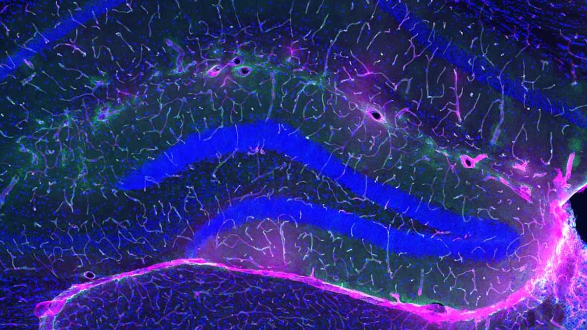
The team used their approach in brain areas known to support different kinds of memory: the dentate gyrus of the hippocampus, which is important for learning and recent recall, and the medial prefrontal cortex, which contributes to remote recall two weeks later.

In aged mice, briefly activating OSK in learning-related hippocampal engram neurons restored memory, essentially bringing performance back to levels seen in young controls. When the same approach was applied to prefrontal cortex engrams, it also recovered remote memories formed weeks earlier.

The reprogrammed engram neurons also showed signs of improved health. They maintained their neuronal identity and displayed molecular features associated with a younger state, including changes in nuclear structure linked to aging.

The team then tested mouse models of Alzheimer’s disease. In a spatial-learning task, the mice showed inefficient navigation and impaired memory strategies. Reprogramming dentate gyrus engrams improved learning strategies during training, while targeting prefrontal engrams restored long-term spatial memory.

Abstract: Stressing the details in the link between chronic stress and liver cancer…

Here, Xuetian Yue discover chronic stress promotes aminopeptidase N expression to increase glutathione synthesis and inhibit ferroptosis in models of liver cancer.


1Department of Cellular Biology, School of Basic Medical Sciences;

2Key Laboratory for Experimental Teratology of Ministry of Education and Department of Immunology, School of Basic Medical Sciences; and.

3Key Laboratory for Experimental Teratology of the Ministry of Education, Department of Histology and Embryology, School of Basic Medical Sciences, Cheeloo Medical College, Shandong University, Jinan, China.

Abstract: Opening the gates to the blood brain barrier (BBB) in a high-grade glioma model

Amy B. Heimberger find therapeutic benefit in adding the STING agonist 8,803 to radiation in preclinical models of glioma. The combination reprogramed the glioma tumor microenvironment, and 8,803 induced the opening of the blood-brain barrier.


3Department of Radiology.

4Department of Neurology, and.

5Department of Radiation Oncology, Feinberg School of Medicine, Northwestern University, Chicago, Illinois, USA.

Tumour–brain crosstalk restrains cancer immunity via a sensory–sympathetic axis

Tumor–brain crosstalk worsens lung cancer.

It is not clear how the brain senses and responds to tumors in peripheral organs, although tumors are innervated by different branches of the peripheral nervous system and increased tumor innervation is associated with poor cancer outcomes.

Authors in this study identify an immuno-suppressive tumor microenvironment established by a tumor–brain axis that promotes oncogenesis.

The researchers demonstrate that lung adenocarcinoma induces innervation and functional engagement of vagal sensory neurons. Mechanistically, the vagal sensory nerves transmit signals from lung tumors to brainstem nuclei, driving elevated sympathetic efferent activity in the tumor microenvironment. This, in turn, suppresses β2 adrenergic signalling in alveolar macrophage and anti-tumor immunity.

Disruption of this sensory-to-sympathetic pathway significantly inhibited lung tumor growth by enhancing immune responses against cancer. sciencenewshighlights sciencemissionn https://sciencemission.com/Tumour%E2%80%93brain-crosstalk


Mouse models demonstrate that vagal sensory neurons transmit signals from lung adenocarcinoma to the brain, increasing sympathetic efferent activity in the tumour microenvironment and thereby creating a immunologically permissive environment for tumour growth.

How dietary restriction rewires immunity to protect against infection

To understand the complex interactions involved in an immune response during scarcity, the team put mice on a 50% restricted-calorie diet and then exposed the animals to bacteria that infect the gut. The mice that were fed a standard diet experienced a metabolic crash— their blood glucose levels and body weight plummeted.

The researchers had expected this would happen to all the animals because mounting an immune response can consume up to 30% of the entire body’s fuel reserves. But in the calorie-restricted mice, the immune system appeared to be functioning perfectly well without using much glucose.

To unravel this enigma, the researchers inventoried the immune cells of the infected animals and discovered that T cells, which normally target invading microbes, were depleted in the calorie-restricted mice. Instead, short-lived neutrophils, which serve as the body’s first responders to infection, were ramped up to twice the normal amount and had measurably enhanced pathogen-killing abilities. The cells seemed to be operating in energy-saving mode, consuming much less glucose than neutrophils from well-fed animals.

The researchers are breaking new ground by outlining how a sudden fall in food intake triggers glucocorticoid levels to rise, resulting in two major shifts. First, the body repositions certain immune cells—especially naïve T cells—into the bone marrow, which becomes a kind of “safe house” for when the cells are needed. Second, during an infection, glucocorticoids tilt the immune response away from energy-intensive T cells toward neutrophils, abundant cells that act as immediate, short-lived defenders.

Beyond clearing a current infection, glucocorticoids prepare the immune system for repeat encounters with infectious agents. While the hormones direct killer T cells to stand down and neutrophils to step up, they also ensure memory T cells are preserved for future confrontations.


When food is scarce, stress hormones direct the immune system to operate in “low power” mode to preserve immune function while conserving energy, according to researchers. This reconfiguration is crucial to combating infections amid food insecurity.

NAD+ sensing by PARP7 regulates the C/EBPβ-dependent transcription program during adipogenesis

Stokes et al. demonstrate that PARP7 “senses” the levels of nuclear NAD+ during early adipogenic differentiation via an ADP-ribosylation-ubiquitin-proteasome pathway to regulate C/EBPβ-dependent proadipogenic gene expression through p300-mediated H3K27 acetylation. Stabilized PARP7 promotes the binding of C/EBPβ to chromatin genome-wide, enhancing lipid synthesis and adipogenesis in vivo.

Genetic defect that weakens esophageal lining identified!

But the molecular factors responsible for the onset of Barrett’s esophagus remain poorly understood.

The findings, published in Nature Communications, combined family studies, laboratory experiments and genetically engineered mouse models to identify and understand how genetic defects contribute to disease development.

The team sequenced and analyzed genetic material of 684 people from 302 families where multiple members developed Barrett’s esophagus or esophageal cancer. They discovered that a subset of affected family members carry inherited mutations in a gene called VSIG10L.

“We found that this gene acts like a quality control system for the esophageal lining,” said the lead researcher. “When it’s defective, the cells do not mature properly and the protective barrier in the esophageal lining becomes weak, allowing stomach bile acid to cause tissue changes that enhances the risk of developing Barrett’s esophagus.”

When researchers genetically engineered mice with human-equivalent VSIG10L mutations, they found that the esophageal lining became disrupted structurally and molecularly, according to the author. The study found that when the mice were exposed to bile acid, they developed Barrett’s-like disease over time, effectively replicating the disease’s progression in humans.

These genetically engineered mice also represent the first animal model for Barrett’s esophagus based directly on human genetic predisposition to the disease, the author said.

With VSIG10L shown to be a key gene in maintaining esophageal health, family members can now be screened for genetic variants to identify those at a high-risk of developing Barrett’s esophagus or esophageal cancer. ScienceMission sciencenewshighlights.

System isolates single extracellular vesicle surface proteins to map function

Extracellular vesicles (EVs) are tiny biological bubbles that carry nucleic acids and proteins between cells, playing an essential role in tissue repair, neuroprotection and immune health. By isolating the surface proteins of these bubbles, researchers can understand more about their biology and build tools to transform extracellular vesicles into next-generation drugs for cancer, neurological conditions and other diseases.

UC Davis biomedical engineers are using EVs to crack the code of the body’s message system. Their findings are detailed in a paper published in ACS Nano.

“EV-mediated intercellular communication is a very powerful system that controls many physiological and pathophysiological phenomena,” said Aijun Wang, a corresponding author of the new study. Wang is Chancellor’s Fellow and professor of biomedical engineering and surgery. “We know that EVs are therapeutically useful. But how do we define what dictates their functions?”

/* */