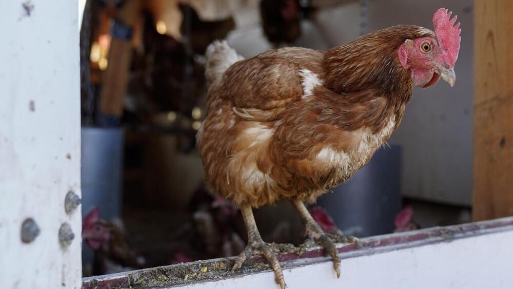
Bats (Mammalia: Chiroptera) serve as natural reservoirs for many zoonotic pathogens worldwide, including vector-borne pathogens. However, bat-associated parasitic arthropods and their microbiota are thus far not thoroughly described in many regions across the globe, nor is their role in the spillover of pathogens to other vertebrate species well understood. Basic epidemiological research is needed to disentangle the complex ecological interactions among bats, their specific ectoparasites and microorganisms they harbor. Some countries, such as Ukraine, are particularly data-deficient in this respect as the ectoparasitic fauna is poorly documented there and has never been screened for the presence of medically important microorganisms. Therefore, the aims of this study were to provide first data on this topic.
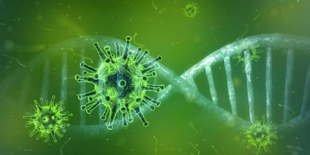
A total of 239 arthropod specimens were collected from bats. They belonged to several major groups of external parasites, including soft ticks, fleas, and nycteribiid flies from six chiropteran species in Northeastern Ukraine. The ectoparasites were individually screened for the presence of DNA of Rickettsia spp., Anaplasma/Ehrlichia spp., Bartonella spp., Borrelia spp., and Babesia spp. with conventional PCRs. Positive samples were amplified at several loci, sequenced for species identification, and subjected to phylogenetic analysis.
Rickettsia DNA was detected exclusively in specimens of the soft tick, Carios vespertilionis (7 out of 43 or 16.3%). Sequencing and phylogenetic analysis revealed high similarity to sequences from Rickettsia parkeri and several other Rickettsia species. Bacteria from the family Anaplasma taceae were detected in all groups of the ectoparasites (51%, 122/239 samples), belonging to the genera Anaplasma, Ehrlichia, and Wolbachia. The detection of Bartonella spp. was successful only in fleas (Nycteridopsylla eusarca) and bat flies (Nycteribia koleantii, N. pedicularia), representing 12.1% (29÷239) of the collected ectoparasites. No DNA of Babesia or Borrelia species was identified in the samples.