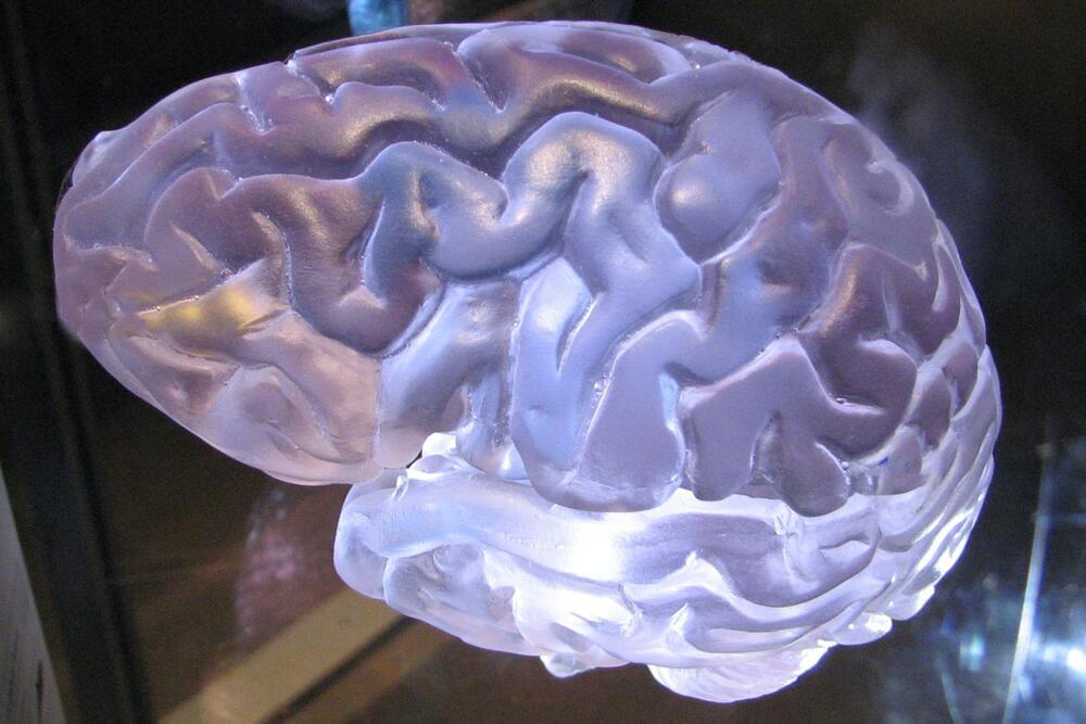
Toggle light / dark theme

Erectile dysfunction drugs may be able to boost the effect of chemotherapy in esophageal cancer

A group of drugs commonly used to treat erectile dysfunction may be able to boost the effect of chemotherapy in esophageal cancer, according to new research funded by Cancer Research UK and the Medical Research Council.

This research, published today (Tuesday) in Cell Reports Medicine, found that the drugs, known as PDE5 inhibitors can reverse chemotherapy resistance by targeting cells called cancer-associated fibroblasts (CAFs) residing in the area surrounding the tumor.

Although this is early discovery research, PDE5 inhibitors combined with chemotherapy may be able to shrink some esophageal tumors more than chemotherapy could alone, tackling chemotherapy resistance, which is one of the major challenges in treating esophageal cancer.

Light-activated “photoimmunotherapy” kills brain cancer, reduces relapse

Scientists at the Institute of Cancer Research in London have developed a new light-activated “photoimmunotherapy” that could help treat brain cancer. The key is a compound that glows under light to guide surgeons to the tumor, while near-infrared light activates a cancer-killing mechanism.

The new study builds on a common technique called Fluorescence Guided Surgery (FGS), which involves introducing a fluorescent agent to the body which glows under exposure to light. This is paired with a synthetic molecule that binds to a specific protein, such as those expressed by cancer cells. The end result is tumors that glow under certain lighting conditions or imaging, guiding surgeons to remove the affected cells more precisely.

For the new study, the researchers gave the technique an extra ability – killing the cancer as well. They added a new molecule that binds to a protein called EGFR, which is often mutated in cases of the brain cancer glioblastoma. After the fluorescence has helped surgeons remove the bulk of the tumor, they can shine near-infrared light on the site, which switches the compound into a tumor-killing mode by releasing reactive oxygen species. The idea is to kill off any remaining cells that could – and often do – stage an aggressive comeback after surgery.

Speeding Up Molecule Design With a New Technique That Can Delete Single Atoms

University of ChicagoFounded in 1,890, the University of Chicago (UChicago, U of C, or Chicago) is a private research university in Chicago, Illinois. Located on a 217-acre campus in Chicago’s Hyde Park neighborhood, near Lake Michigan, the school holds top-ten positions in various national and international rankings. UChicago is also well known for its professional schools: Pritzker School of Medicine, Booth School of Business, Law School, School of Social Service Administration, Harris School of Public Policy Studies, Divinity School and the Graham School of Continuing Liberal and Professional Studies, and Pritzker School of Molecular Engineering.

Patients Treated With CRISPR Still “Cured” Three Years Later

A long-running human trial has shown that CRISPR gene editing could prove to be a highly effective way of treating serious conditions.

The trial, which was kicked off in 2019 by an international team of scientists, found that a new gene-editing therapy called exagamglogene autotemcel, or ex-cel for short, was able to essentially “cure” patients with transfusion-dependent beta thalassemia (TDT) or severe sickle cell disease (SCD), two blood disorders that are conventionally treated using blood transfusions.

It’s a promising new use of the technology. Around 100,000 Americans are affected by TDT, while SCD affects an estimated 300 to 3,000. And in a broader sense, the results suggest that tinkering with genetic code could come to be a practical, widespread new area of medicine.

Open-source program IDs synthetic, naturally occurring gene sequences

It’s a given that certain bacteria and viruses can cause illness and disease, but the real culprits are the sequences of concern that lie within the genomes of these microbes.

Calling them out is about to get easier.

Years of work by Rice University computer scientists and their colleagues have led to an improved platform for DNA screening and pathogenic sequence characterization, whether naturally occurring or synthetic, before they have the chance to impact public health.

Biochemists use new tool to control mRNA

A team of researchers at the Institute of Biochemistry at Münster University discovered that by using so-called FlashCaps they were able to control the translation of mRNA by means of light. The results have been published in Nature Chemistry.

DNA () is a long chain of molecules composed of many individual components, and it forms the basis of life on Earth. The function of DNA is to store all genetic information. The translation of this into proteins—which an organism needs to function, develop and reproduce—takes place via mRNA (messenger ribonucleic acid). The DNA is transcribed to mRNA, and the mRNA in turn is translated into proteins (protein biosynthesis). In other words, the mRNA functions as an information carrier. Biochemists at the University of Münster have now developed a new biochemical tool that is able to to control the translation of RNA with the aid of light. These so-called FlashCaps enable researchers to control a variety of processes in cells both spatially and temporally and, as a result, to determine basic functions of proteins.

Switching DNA functions on and off with light

DNA is the basis of life on earth. The function of DNA is to store all the genetic information an organism needs to develop, function and reproduce. It is essentially a biological instruction manual found in every cell. Biochemists at the University of Münster have now developed a strategy for controlling the biological functions of DNA with the aid of light. This enables researchers to better understand and control the processes that take place in the cell—for example, epigenetics, the key chemical change and regulatory lever in DNA. The results have been published in the journal Angewandte Chemie.

The cell’s functions depend on enzymes. Enzymes are proteins that carry out in the cell. They help to synthesize metabolic products, make copies of the DNA molecules, convert energy for the cell’s activities, change DNA epigenetically and break down certain molecules. A team of researchers headed by Prof. Andrea Rentmeister from the Institute of Biochemistry at the University of Münster used a so-called enzymatic cascade reaction to understand and track these functions better. This sequence of successive reaction steps involving different enzymes makes it possible to transfer so-called photocaging groups—chemical groups that can be removed by means of irradiation with light—to DNA. Previously, studies had shown that only small residues (small modifications such as methyl groups) could be transferred selectively to DNA, RNA (ribonucleic acid) or proteins.

“As a result of our work, it is now possible to transfer larger residues or modifications such as the photocaging groups just mentioned,” explains Nils Klöcker, one of the lead authors of the study and a Ph.D. student at the Institute of Biochemistry. Working together with structural biologist Prof. Daniel Kümmel, who also works at the Institute of Biochemistry, it was also possible to explain the basis for the changed activity at a .

A neural autoencoder to enhance sensory neuroprostheses

New technologies have the potential to greatly simplify the lives of humans, including those of blind individuals. One of the most promising types of tools designed to assist the blind are visual prostheses.

Visual prostheses are that can be implanted in the brain. These devices could help to restore vision in people affected by different types of blindness. Despite their huge potential, most existing visual prostheses achieved unimpressive results, as the vision they can produce is extremely rudimentary.

A team of researchers a University of California, Santa Barbara recently developed a that could significantly enhance the performance of visual prostheses, as well as other sensory neuroprostheses (i.e., devices aimed at restoring lost sensory functions or augmenting human abilities). The model they developed, presented in a paper pre-published on arXiv, is based on the use of a neural autoencoder, a brain-inspired architecture that can discover specific patterns in data and create representations of them.

Functional DNA-based cytoskeletons for synthetic cells

Cytoskeletons are essential components of cells that perform a variety of tasks, and artificial cytoskeletons that perform these functions are required for the bottom-up assembly of synthetic cells. Now, a multi-functional cytoskeleton mimic has been engineered from DNA, consisting of confined DNA filaments that are capable of reversible self-assembly and transport of gold nanoparticles and vesicular cargo.

/* */