Year 2022 This is their published work on the universal vaccine of the covid 19.
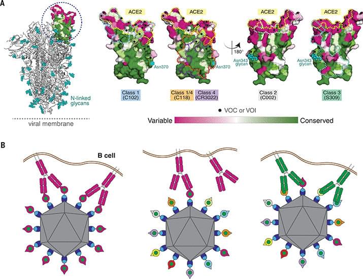
A mosaic sarbecovirus nanoparticle protects against SARS-2 and SARS-1, whereas a SARS-2 nanoparticle only protects against SARS-2.
(NewsNation) — Studies have shown that Alzheimer’s may become the defining disease of the baby boomer generation.
According to The Alzheimer’s Association, the number of people age 65 and over living with Alzheimer’s now is nearly 7 million. That number is expected to rise to over 13 million by 2050.
Physician and best-selling author Dr. Ian Smith says it’s not known exactly what causes Alzheimer’s.
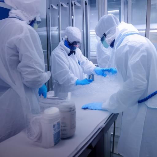
What if death was not the end? What if, instead of saying our final goodbyes to loved ones, we could freeze their bodies and bring them back to life once medical technology has advanced enough to cure their fatal illnesses? This is the mission of Tomorrow Biostasis, a Berlin-based startup that specializes in cryopreservation.
Cryopreservation, also known as biostasis or cryonics, is the process of preserving a human body (or brain) in a state of suspended animation, with the hope that it can be revived in the future when medical technology has advanced enough to treat the original cause of death. This may seem like science fiction, but it is a legitimate scientific procedure, and Tomorrow Biostasis is one of the few companies in the world that offers this service.
Dr Emil Kendziorra, co-founder and CEO of Tomorrow Biostasis explained that the goal of cryopreservation is to extend life by preserving the body until a cure can be found for the original illness. He emphasized that cryopreservation is not a form of immortality, but rather a way to give people a second chance at life.
Increasingly dense cell clusters in growing tumors convert blood vessels into fiber-filled channels. This makes immune cells less effective, as findings by researchers from ETH Zurich and the University of Strasbourg suggest. Their research is published in Matrix Biology.
It was almost ten years ago that researchers first observed that tumors occurring in different cancers—including colorectal cancer, breast cancer and melanoma—exhibit channels leading from the surface to the inside of the cell cluster. But how these channels form, and what functions they perform, long remained a mystery.
Through a series of elaborate and detailed experiments, the research groups led by Viola Vogel, Professor of Applied Mechanobiology at ETH Zurich, and Gertraud Orend from the University of Strasbourg have found possible answers to these questions. There is a great deal of evidence to suggest that these channels, which the researchers have dubbed tumor tracks, were once blood vessels.
Brainoids — tiny clumps of human brain cells — are being turned into living artificial intelligence machines, capable of carrying out tasks like solving complex equations. The team finds out how these brain organoids compare to normal computer-based AIs, and they explore the ethics of it all.
Sickle cell disease is now curable, thanks to a pioneering trial with CRISPR gene editing. The team shares the story of a woman whose life has been transformed by the treatment.
We can now hear the sound of the afterglow of the big bang, the radiation in the universe known as the cosmic microwave background. The team shares the eerie piece that has been transposed for human ears, named by researchers The Echo of Eternity.
Human Cyborg — We’ve all seen Cyborgs in Hollywood blockbusters. But it turns out these fictional beings aren’t so far-fetched.
Human Cyborg (2020)
Director: Jacquelyn Marker.
Writers: Kyle McCabe, Christopher Webb Young.
Stars: Justin Abernethy, Robert Armiger, John Donoghue.
Genre: Documentary.
Country: United States.
Language: English.
Also Known As: Cyborg Revolution.
Release Date: 2020 (United States)
Synopsis:
We’ve all seen Cyborgs in Hollywood blockbusters. But it turns out these fictional beings aren’t so far-fetched. In fact, this episode features a true-to-life cyborg, who at four months of age, was the youngest American to be outfitted with a myoelectric hand. And at one ground-breaking engineering facility, engineers are developing biotechnologies that can even further enhance high-tech like this by giving mechanical prosthetics something incredible: the physical sensation of touch!
Other engineering firms are gearing up powerful exoskeletons that both rehabilitate and enhance the power of the human body… improving the lives of those with paralysis and transforming the work force.
But the real pivot is getting machines inside the body. An out-of-the-box ‘transhumanist’ featured in the episode installs a chip inside a person’s hand. It works as a key that unlocks doors, literally and figuratively.
However, brain-machine integration poses the biggest challenges, and the biggest rewards. Cutting-edge neuroscientists and technologists reveal how computer chips can directly interface with the human brain in ways that not only rehabilitate, but which can also ‘read thoughts’ in real-time.

The Maerdang plant will have a total installed capacity of around 2.2 million kW.
In an effort to ramp up its renewable energy production, China is on course to begin operations of its highest-altitude hydropower.
A clean energy initiative to optimize resources
The Maerdang project will feature an integrated clean energy approach that includes hydropower, solar power, and energy storage. The project serves as an example of how China is leveraging its clean energy sources in its western regions to supply the growing national demand for energy.
“The company vows to make sure the project is put into operation in time despite COVID-19 impacts during the past few years,” said Li Hongxin, deputy director of Qinghai Maerdang Co Ltd, a unit of China Energy, told China Daily.
The research team used a new CRISPR-based genome editing system named PESpRY.
Scientists in China have effectively treated retinitis pigmentosa.
The research team utilized a novel form of CRISPR-based genome editing that is exceptionally adaptable and could potentially remedy numerous genetic mutations responsible for causing different diseases.