Here’s my new Opinion essay at Newsweek. It’s about the need to use our nation’s massive natural resources to pay for a bipartisan tax free universal basic income, called the Federal Land Dividend. I hope you will read and share it!
In 2018, I began lecturing about the Federal Land Dividend, a bipartisan tax-free Universal Basic Income (UBI) based on monetizing the 640 million acres of mostly unused federally owned land. Due to the lasting effects of the coronavirus pandemic, which include a struggling U.S. economy, there is increasing interest in implementing basic income plans. The Federal Land Dividend is the only method that is both bipartisan and tax free.
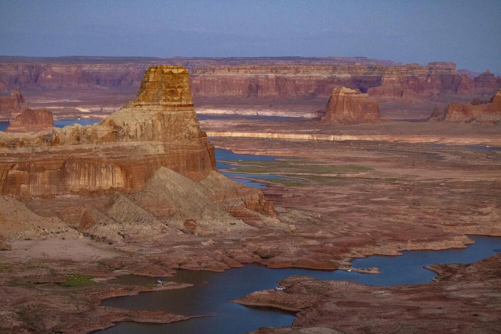
An estimated 50 percent of the 11 most western states are mostly empty land that belong to the government. Estimates say this land and its resources are worth approximately $100 to $200 trillion. If we divide the middle— $150 trillion —by America’s population of 333 million, every person would have approximately $450,000 in equity. That’s much higher than the median net worth in America of $122,000.
The Federal Land Dividend aims to lease out land and natural resources to big business that agree, in exchange, to pay a monthly income to all Americans. It’s estimated that if just 60 percent of America’s unused federal land was leased out at fair rates, a $1,000 monthly check could be sent to all Americans—regardless of age—for decades if not centuries. Because land and raw materials often move in tandem with inflation, payouts could increase with inflation. Furthermore, this plan does not touch any national parks whatsoever. Much of this land is in places that few humans ever visit or see.
Some conservatives support the Federal Land Dividend idea because it will boost big business while providing an economic stimulus to all Americans. Some liberals also support the idea because it will dramatically help end poverty. Even some libertarians like the idea because it returns the value of federal land to the people, instead of the government hoarding and controlling it.