The Nobel Awards Season just ended, with the “Oscars of Science” awarded to some of the world’s brightest minds. The entire science world was watching, and just like with the Oscars, there was an element of suspense, drama, envy, celebration, and happiness. Most of the Nobel Laureates are also phenomenal speakers and communicators with decades of teaching experience, and thousands of people across the world are glued to their monitors to hear their inspiring stories. The Nobel Prizes are awarded in Physics, Chemistry, Physiology or Medicine, Literature, Peace, and Economic Sciences. Unfortunately, there is no Nobel Prize for Computer Science, Mathematics, or Engineering. So, it seems like it… More.
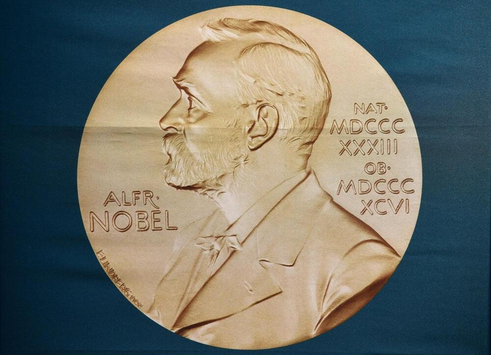
While there is no Nobel Prize for AI, Jumper and Hassabis may be the frontrunners for a Nobel Prize in Chemistry for their discovery of AlphaFold.