Scientists introduce Zman-seq, a method revolutionizing our understanding of dynamic cellular changes in the human body over time. Read more about this groundbreaking study.
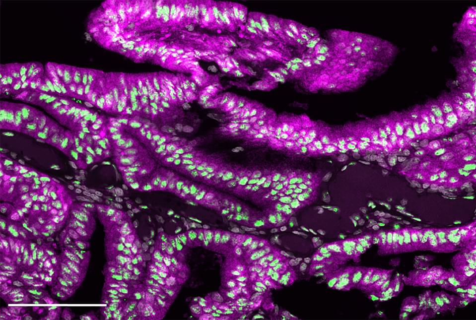
In a recent study published in Cell, scientists led by Prof. Ido Amit at the Weizmann Institute of Science have introduced Zman-seq. This revolutionary method breaks through the temporal barriers of cellular analysis.
This innovative approach allows tracking and measuring changes in individual cells within the body over time.
“Knowing what preceded what is not enough to deduce causality, but without this knowledge, we don’t have a chance of understanding what the cause and effect are,” said Prof. Amit in a press release.