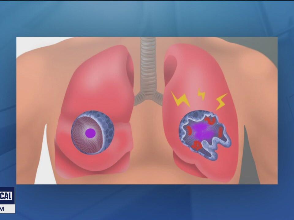
St. Jude Children’s Research Hospital scientists reversed an aggressive cancer, reverting malignant cells towards a more normal state. Rhabdoid tumors are an aggressive cancer which is missing a key tumor suppressor protein. Findings showed that with the missing tumor suppressor, deleting or degrading the quality control protein DCAF5 reversed the cancer cell state. These results suggest a new approach to curing cancer — returning cancerous cells to an earlier, more normal state rather than killing cancer cells with toxic therapies — may be possible. The results were published today in Nature.
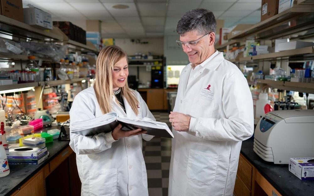
“Rather than making a toxic event that kills rhabdoid cancer, we were able to reverse the cancer state by returning the cells toward normal,” said senior author Charles W.M. Roberts, MD, PhD, Executive Vice President and St. Jude Comprehensive Cancer Center director. “This approach would be ideal, especially if this paradigm could also be applied to other cancers.”
“We found a dependency which actually reverses the cancer state,” said first author Sandi Radko-Juettner, PhD, a former St. Jude Graduate School of Biomedical Sciences student, now a Research Program Manager for the Hematological Malignancies Program at St. Jude. “Standard cancer therapies work by causing toxicities that also damage healthy cells in the body. Here, it appears that we’re instead fixing the problem caused by the loss of a tumor suppressor in this rhabdoid cancer.”