Toggle light / dark theme

Utilizing neurotransmitters as a passport into the brain:


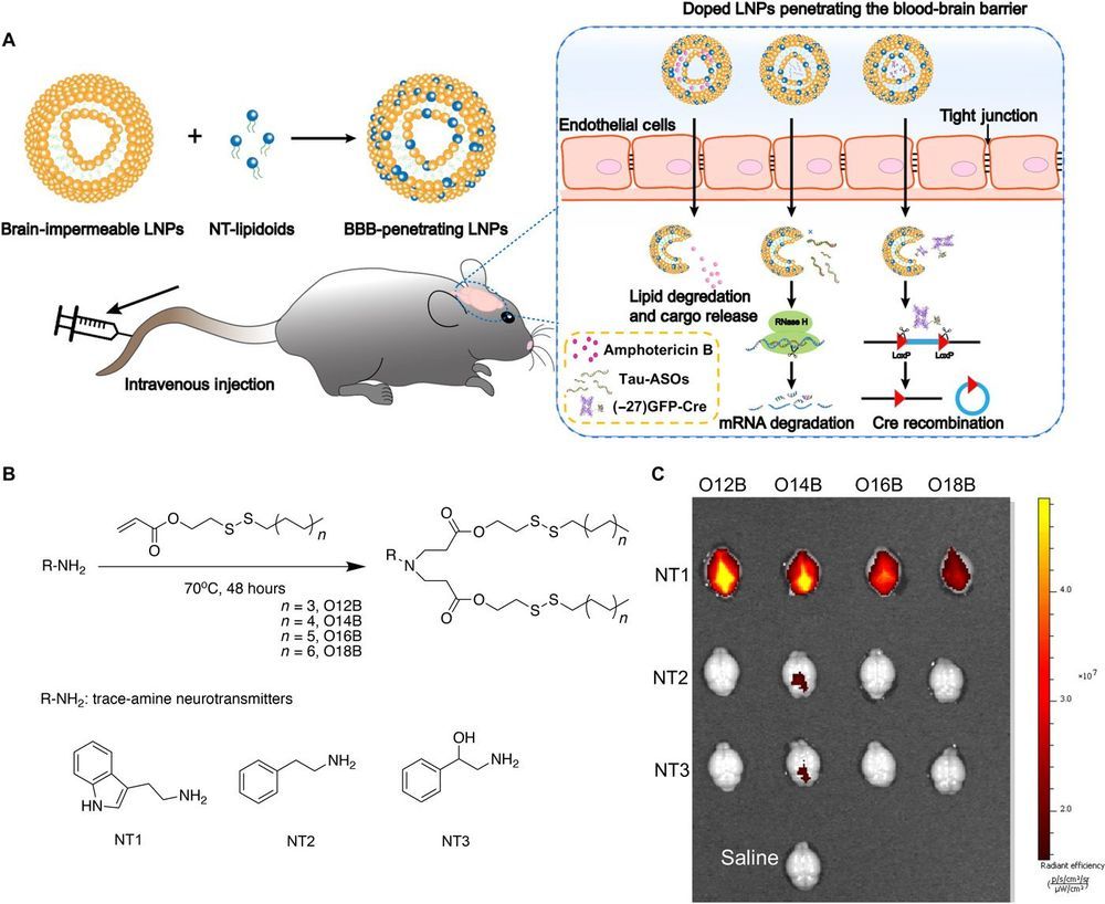
Safe and efficient delivery of blood-brain barrier (BBB)–impermeable cargos into the brain through intravenous injection remains a challenge. Here, we developed a previously unknown class of neurotransmitter–derived lipidoids (NT-lipidoids) as simple and effective carriers for enhanced brain delivery of several BBB-impermeable cargos. Doping the NT-lipidoids into BBB-impermeable lipid nanoparticles (LNPs) gave the LNPs the ability to cross the BBB. Using this brain delivery platform, we successfully delivered amphotericin B (AmB), antisense oligonucleotides (ASOs) against tau, and genome-editing fusion protein (−27)GFP-Cre recombinase into the mouse brain via systemic intravenous administration. We demonstrated that the NT-lipidoid formulation not only facilitates cargo crossing of the BBB, but also delivery of the cargo into neuronal cells for functional gene silencing or gene recombination. This class of brain delivery lipid formulations holds great potential in the treatment of central nervous system diseases or as a tool to study the brain function.
Objective To determine whether training with a brain–computer interface (BCI) to control an image of a phantom hand, which moves based on cortical currents estimated from magnetoencephalographic signals, reduces phantom limb pain.

Methods Twelve patients with chronic phantom limb pain of the upper limb due to amputation or brachial plexus root avulsion participated in a randomized single-blinded crossover trial. Patients were trained to move the virtual hand image controlled by the BCI with a real decoder, which was constructed to classify intact hand movements from motor cortical currents, by moving their phantom hands for 3 days (“real training”). Pain was evaluated using a visual analogue scale (VAS) before and after training, and at follow-up for an additional 16 days. As a control, patients engaged in the training with the same hand image controlled by randomly changing values (“random training”). The 2 trainings were randomly assigned to the patients. This trial is registered at UMIN-CTR (UMIN000013608).

Results VAS at day 4 was significantly reduced from the baseline after real training (mean [SD], 45.3 [24.2]–30.9 [20.6], 1/100 mm; p = 0.009 < 0.025), but not after random training (p = 0.047 0.025). Compared to VAS at day 1, VAS at days 4 and 8 was significantly reduced by 32% and 36%, respectively, after real training and was significantly lower than VAS after random training (p < 0.01).

When our neurons—the principle cells of the brain—die, so do we.

Most neurons are created during and have no “backup” after birth. Researchers have generally believed that their survival is determined nearly extrinsically, or by outside forces, such as the tissues and that neurons supply with .

A research team led by Sika Zheng, a biomedical scientist at the University of California, Riverside, has challenged this notion and reports the continuous survival of neurons is also intrinsically programmed during development.

Even healthy brains become less efficient as they age, but they do so at different rates for different tasks in different people. Understanding what contributes to this decline, and the ways in which that decline varies, can provide significant insight into the function of the brain.

In a new study, researchers at The University of Texas at Dallas documented how some parts of the brain perform differently over time in response to various kinds of visual input.

A team from the Center for Vital Longevity (CVL) analyzed a phenomenon called neural dedifferentiation, in which regions of the brain that normally are specialized to perform distinct tasks become less selective in their responses to stimulus types.

The thalamus is a “Grand Central Station” for sensory information coming to our brains. Almost every sight, sound, taste and touch we perceive travels to our brain’s cortex via the thalamus. It is theorized that the thalamus plays a major role in consciousness itself. Not only does sensory information pass through the thalamus, it is also processed and transformed by the thalamus so our cortex can better understand and interpret these signals from the world around us.

One powerful type of transformation comes from interactions between excitatory neurons that carry data to the neocortex and inhibitory neurons of the thalamic reticular nucleus, or TRN, that regulate flow of that data. Although the TRN has long been recognized as important, much less has been known about what kinds of cells are in the TRN, how they are organized and how they function.

Now a paper published in the journal Nature addresses those questions. Researchers led by corresponding author Scott Cruikshank, Ph.D., and co-authors Rosa I. Martinez-Garcia, Ph.D., Bettina Voelcker, Ph.D., and Barry Connors, Ph.D., show that the somatosensory part of the TRN is divided into two functionally distinct sub-circuits. Each has its own types of genetically defined neurons that are topographically segregated, are physiologically distinct and connect reciprocally with independent thalamocortical nuclei via dynamically divergent synapses.

Goodbye depression.


Neuralink‘s mission has never quite been clear. We know it’s working on a chip designed to be surgically inserted into the human skull called a brain-computer interface (BCI), but exactly what and who it’s for remains a bit of a mystery.

As best we can tell based on what’s been revealed so far, it’s shaping up to be a terrifying hormone hijacker capable of potentially giving you forced mental orgasms or making you fall in love.

Yes.

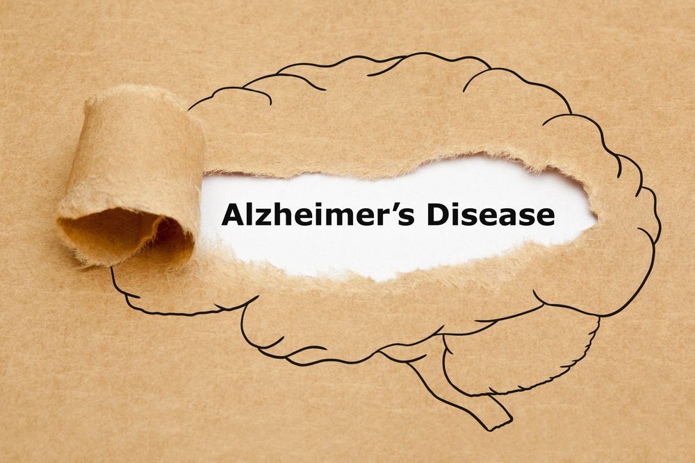
TOKYO – Although scientists know many of the underlying symptoms which trigger Alzheimer’s disease, a cure remains elusive. Now, a new study suggests that oxytocin, a hormone best known for promoting feelings of love and wellbeing, may reverse some of the damage the degenerative illness causes.

Alzheimer’s disease is a progressive brain disease causing the continuous deterioration of mental functions. Its primary symptoms include severely impaired thinking, memory loss, and confusion.

One of the primary culprits in Alzheimer’s is a protein known as amyloid β (Aβ). Researchers say Aβ clumps together to form plaques around neurons in the brain. These plaque build-ups disrupt normal neuron function and triggers the degeneration.