Toggle light / dark theme

Join us on Patreon! https://www.patreon.com/MichaelLustgartenPhD

Discount Links/Affiliates:
Blood testing (where I get my labs): https://www.ultalabtests.com/partners/michaellustgarten.

Clearly Filtered Water Filter: https://get.aspr.app/SHoPY

At-Home Metabolomics: https://www.iollo.com?ref=michael-lustgarten.

This member-only story is on us. Upgrade to access all of Medium.

Now that AI is transforming nearly every industry, healthcare stands out as a field with immense potential — and unique risks.

A single AI-generated error here could lead to serious consequences for patient health.

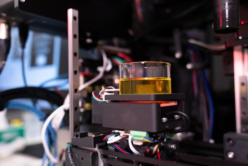
Biomedical engineers from the University of Melbourne have invented a 3D printing system, or bioprinter, capable of fabricating structures that closely mimic the diverse tissues in the human body, from soft brain tissue to harder materials like cartilage and bone.

This cutting-edge technology offers cancer researchers an advanced tool for replicating specific organs and tissues, significantly improving the potential to predict and develop new pharmaceutical therapies. This would pave the way for more advanced and ethical drug discovery by reducing the need for animal testing.

Head of the Collins BioMicrosystems Laboratory at the University of Melbourne, Associate Professor David Collins said: In addition to drastically improving print speed, our approach enables a degree of cell positioning within printed tissues. Incorrect cell positioning is a big reason most 3D bioprinters fail to produce structures that accurately represent human tissue.

Within the body, cells involved in specific functions, like immune response and secretion, are equipped with granules, which are small membrane-bound compartments containing enzymes, proteins, or other molecules. In neutrophils, the so-called azurophilic (or primary) granules contain enzymes that are involved in the initial response to an infection.

MPO-based E-101 is the first wound and systemically safe antiseptic, with a safety profile comparable to saline, that is effective in physiological conditions.

Myeloperoxidase, or MPO, is one of the most important of such enzymes for the immune system’s ability to destroy pathogens. Allen has been studying the physiological role of MPO since 1971. Using chemiluminescence and metabolic studies he has been able to study the complex and finely regulated mechanism of NADPH oxidase driven MPO action in microbicidal activity.

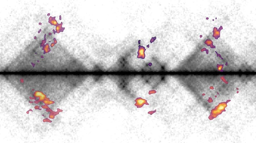
New research from the Kind Group at the Hubrecht Institute sheds light on how cells repair damaged DNA. For the first time, the team has mapped the activity of repair proteins in individual human cells.

The study demonstrates how these proteins collaborate in so-called “hubs” to repair DNA damage. This knowledge offers opportunities to improve cancer therapies and other treatments where DNA repair is essential. The researchers published their findings in Nature Communications on November 21.

DNA is the molecule that carries our genetic information. It can be damaged by normal cellular processes as well as external factors such as UV radiation and chemicals. Such damage can lead to breaks in the DNA strand. If DNA damage is not properly repaired, mutations can occur, which may result in diseases like cancer. Cells use repair systems to fix this damage, with specialized proteins locating and binding to the damaged regions.

Summary: Researchers have demonstrated how brain activity can predict behavior in urban environments, providing a roadmap for improving urban planning. Using functional MRI scans, the study identified activity in the brain’s reward system, specifically the ventromedial prefrontal cortex, as a key predictor of why people visit certain urban areas.

Participants rated photos of Lisbon’s urban spaces, and their brain responses were linked to visitation patterns, showing that people are drawn to areas of perceived value. This research suggests urban design can prioritize environments that align with cognitive and emotional well-being.

Neurourbanism, the emerging field behind this study, offers tools to design cities that enhance livability and sustainability. By focusing on human-centered approaches, cities can improve efficiency, mobility, and resident happiness.

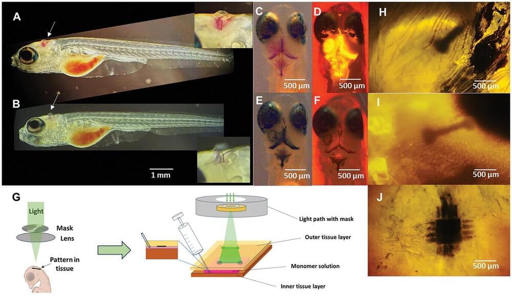
Bioelectronics research and development of implants made of electrically conductive materials for disease treatment is advancing rapidly. However, bioelectronic treatment is not without complications. Researchers at Lund University in Sweden have taken another step forward by developing a refined method to create detailed and tissue-friendly bioelectronics.

In a study published in Advanced Science, the researchers describe how they can use light to create electrically directly in the body, showing promising results in animal trials.

Bioelectronics is successfully used for treating heart arrhythmia, epilepsy, and like Parkinson’s, to name a few. However, it’s well known that today’s bioelectrodes and implantation methods require the tissue to adapt to the electrodes, rather than the other way around. This can lead to complications.

The United States Food and Drug Administration has just approved the first-ever clinical trial that uses CRISPR-Cas13 RNA editing. Its aim is to treat an eye disease called wet age-related macular degeneration that causes vision loss in millions of older people worldwide.

This trial marks a new frontier in —the process of treating or curing medical conditions by changing a person’s genes.

What makes it special is the fact the therapy targets RNA, instead of DNA. So, what does that mean, and why should we be excited?