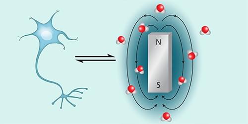
A new technique combining magnetic resonance imaging and x-ray fluorescence can characterize, with single-neuron resolution, the presence of toxic forms of iron that might be associated with neurodegenerative diseases.
Iron plays a major role in life. Most obviously, it keeps us alive, helping to ferry oxygen around our bloodstreams. It is also essential in cellular energy production, in the immune-system response, and in brain function—where it helps catalyze the synthesis of dopamine and other neurotransmitters. Iron can, however, be a double-edged sword. An iron excess has been implicated in many ailments, including neurodegenerative conditions such as Alzheimer’s, multiple sclerosis, and Parkinson’s disease—where dopaminergic neurons (neurons that use iron to synthesize dopamine) degenerate. It is thought that the toxicity of iron depends on how it is stored: iron firmly bound within proteins such as ferritin may be less toxic than iron more loosely bound to low-affinity sites, where it is more able to participate in reactions that generate cell-damaging hydroxyl radicals [1].


Leave a reply