Effort to scan the entire Human Brain continues.
Summary: A new, non-invasive neuroimaging technique allowed researchers to investigate the visual sensory thalamus, a brain area associated with visual difficulties in dyslexia and other disorders.
Source: TU Dresden
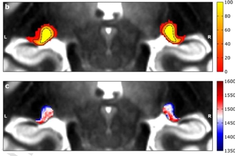
The visual sensory thalamus is a key region that connects the eyes with the cerebral cortex. It contains two major compartments. Symptoms of many diseases are associated with alterations in this region. So far, it has been very difficult to assess these two compartments in living humans, because they are tiny and located very deep inside the brain.
This difficulty of investigating the visual sensory thalamus in detail has hampered the understanding of the function of visual sensory processing tremendously in the past. By coincidence, Christa Müller-Axt, Ph.D. student in the lab of neuroscientist Prof. Katharina von Kriegstein at TU Dresden, discovered structures that she thought might resemble the two visual sensory thalamus compartments in neuroimaging data.


Comments are closed.