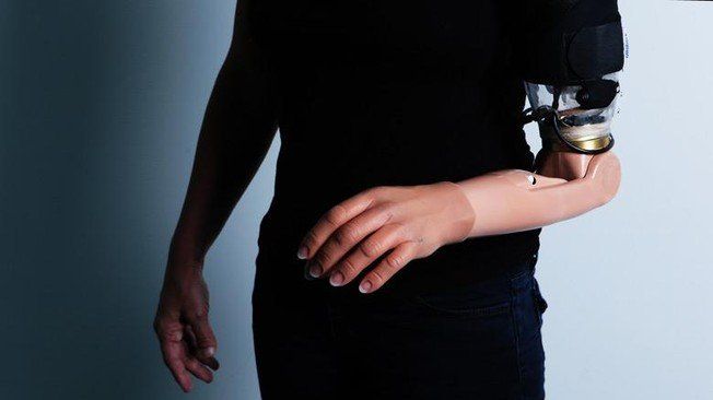
EPFL scientists from the Center for Neuroprosthetics have used functional MRI to show how the brain re-maps motor and sensory pathways following targeted motor and sensory reinnervation (TMSR), a neuroprosthetic approach where residual limb nerves are rerouted towards intact muscles and skin regions to control a robotic limb.
Targeted motor and sensory reinnervation (TMSR) is a surgical procedure on patients with amputations that reroutes residual limb nerves towards intact muscles and skin in order to fit them with a limb prosthesis allowing unprecedented control. By its nature, TMSR changes the way the brain processes motor control and somatosensory input; however the detailed brain mechanisms have never been investigated before and the success of TMSR prostheses will depend on our ability to understand the ways the brain re-maps these pathways. Now, EPFL scientists have used ultra-high field 7 Tesla fMRI to show how TMSR affects upper-limb representations in the brains of patients with amputations, in particular in primary motor cortex and the somatosensory cortex and regions processing more complex brain functions. The findings are published in Brain.
Targeted motor and sensory reinnervation (TMSR) is used to improve the control of upper limb prostheses. Residual nerves from the amputated limb are transferred to reinnervate and activate new muscle targets. This way, a patient fitted with a TMSR prosthetic “sends” motor commands to the re-innervated muscles, where his or her movement intentions are decoded and sent to the prosthetic limb. On the other hand, direct stimulation of the skin over the re-innervated muscles is sent back to the brain, inducing touch perception on the missing limb.


Comments are closed.