I had to take a second review of this since I posted it, and right away I see something quite interesting that folks have overlooked for a while. Will keep you posted.
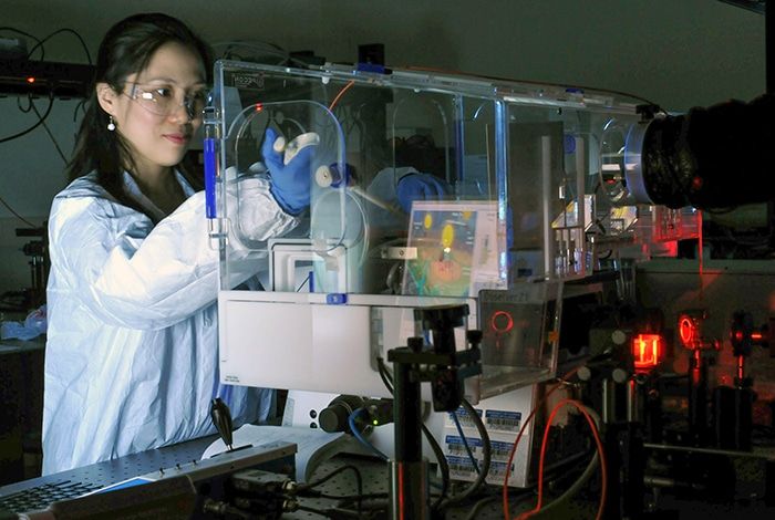
Scientists funded by the National Institutes of Health have built a new tool to monitor the way cells attach to an adjoining substrate under a microscope.
Analyzing adhesion events can help researchers to understand the way diseases spread, tissues grow, and stem cells differentiate into many specific cell types. The technique provides high-resolution images that can monitor the interactions of cells across longer time periods than previously possible.
Researchers from the University of Illinois at Urbana-Champaign published details of their study in the November 2016 issue of the Progress in Quantum Electronics journal. The researchers demonstrated the method known as photonic crystal-enhanced microscopy (PCEM), on various types of cancer cells and stem cells, revealing that each cell type had its own exclusive adhesion properties.


Comments are closed.