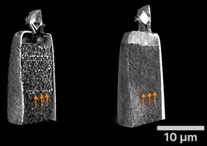
X-ray microscopes are essential for examining components and materials because they can be used to detect changes and details in the material. Until now, however, it has been difficult to detect small cracks or tiny inclusions in the images. By developing a new method, researchers at the Helmholtz-Zentrum Hereon are now able to visualize such changes in the nanometer regime. In particular materials research and quality assurance will profit from this development.
The team reported on their new development in the scientific journal Optica (“Nanoscale dark-field imaging in full-field transmission X-ray microscopy”).
The quality must be right. This also applies to materials science. When metal parts are welded together, you need to know whether the weld seam is any good — or whether small cracks or pores have formed inside, which could lead to failure. High-performance materials, e.g. for electrodes in electric car batteries or fuel cells, should not contain defects to allow the current to flow undisturbed.


